| World Journal of Nephrology and Urology, ISSN 1927-1239 print, 1927-1247 online, Open Access |
| Article copyright, the authors; Journal compilation copyright, World J Nephrol Urol and Elmer Press Inc |
| Journal website https://wjnu.elmerpub.com |
Case Report
Volume 15, Number 4, November 2026, pages 106-112

Endoscopic Management of Full-Thickness Ureteral Perforation Caused by Inadvertent Inflation of a Foley Catheter Balloon Within the Ureter Using Dual Double-J Stenting
Ibrahim Erol 
Department of Urology, Aksaray University Training and Research Hospital, 68100 Aksaray, Turkiye
Manuscript submitted June 25, 2026, accepted September 28, 2026, published online November 18, 2026
Short title: Dual Double-J Stenting for Foley-Related UP
doi: https://doi.org/10.14740/wjnu1063
| Abstract | ▴Top |
Inadvertent inflation of a Foley catheter balloon within the ureter is a rare but potentially severe complication of urethral catheterization. We report a 68-year-old bedridden woman with advanced Alzheimer’s disease, a clinical diagnosis of neurogenic bladder, markedly reduced bladder capacity, and chronic indwelling catheterization who presented 12 h after routine catheter exchange with right flank pain, fever, and gross hematuria. Non-contrast computed tomography (CT) demonstrated a 16 Fr Foley balloon inflated within the right distal ureter immediately below the iliac vessel crossing. Emergency ureteroscopy showed an approximately 2.5-cm full-thickness defect with complete contrast extravasation. The intact lateral ureteral wall was followed to re-enter the true lumen and obtain renal pelvic access. Two 6 Fr double-J stents were selected intraoperatively because of the extensive defect, relatively wide ureteral lumen, and need for reliable internal drainage; the rationale was to improve luminal stability and preserve drainage if one stent migrated or became obstructed. Percutaneous nephrostomy was reserved for failure of retrograde drainage because collecting-system dilatation was minimal. Clinical and laboratory parameters improved after drainage and antibiotic therapy. Postoperative day 2 imaging showed regression of periureteral inflammation without urinary extravasation, and retrograde pyelography at 6 weeks showed ureteral continuity without leakage before stent removal. No subsequent 3- or 6-month imaging was available because the patient did not return for long-term follow-up. Dual double-J stenting may be a technically feasible option in carefully selected extensive injuries when retrograde true-lumen access and adequate drainage can be achieved, but this single case does not establish superiority over single stenting, nephrostomy, or reconstruction.
Keywords: Foley catheter; Ureteral perforation; Double-J stent; Neurogenic bladder; Iatrogenic injury; Ureteroscopy
| Introduction | ▴Top |
Inadvertent ureteral cannulation during urethral catheterization is uncommon but can result in obstruction, infection, acute kidney injury (AKI), or ureteral rupture. An updated literature review of reported cases identified female sex, long-term catheterization, neurologic disease, and altered lower urinary tract anatomy as recurring risk factors [1–6]. Markedly reduced bladder capacity and chronic catheter-related changes may further facilitate entry of a catheter into a ureteral orifice.
Management depends on the extent of injury, the ability to establish urinary drainage, and the patient’s clinical condition. We present a full-thickness distal ureteral perforation caused by Foley balloon inflation in which the true lumen was re-entered endoscopically and two simultaneous double-J stents were used as a selected internal drainage strategy. The case is reported to clarify the intraoperative reasoning for dual stenting and the circumstances in which nephrostomy was retained as a rescue option rather than used initially.
| Case Report | ▴Top |
A 68-year-old bedridden woman with advanced Alzheimer’s disease had a clinical diagnosis of neurogenic bladder; formal urodynamic testing had not been performed. Cystoscopic assessment had demonstrated a markedly reduced bladder capacity of less than 100 mL. Voiding cystourethrography had not been performed, and vesicoureteral reflux (VUR) had therefore not been documented. Following hip fracture surgery in 2023, persistent urinary incontinence contributed to pressure-ulcer care difficulties, and long-term indwelling urethral catheterization was instituted. The catheter was routinely exchanged approximately every 15 days. There was no history of previous urological surgery. A reliable pre-admission medication list was not available in the records reviewed. This report describes a single clinical case and does not constitute systematic research involving human participants. Therefore, ethics committee approval was not required according to local regulations.
Approximately 12 h after a routine catheter exchange, the patient presented with right flank pain, fever, gross hematuria, and impaired oral intake. Oral temperature was 39.0 °C, heart rate 90 beats/min, blood pressure 100/60 mm Hg, respiratory rate 24 breaths/min, and peripheral oxygen saturation 92%. She was hemodynamically stable. Serum lactate was not measured. Laboratory analysis showed leukocytosis (35,000/µL), C-reactive protein (CRP) 180 mg/L, and AKI with serum creatinine 2.01 mg/dL. During bladder irrigation, resistance and insufficient return of irrigation fluid prompted urgent urological assessment.
Non-contrast abdominal computed tomography (CT) demonstrated a 16 Fr Foley catheter balloon fully inflated within the right ureter immediately below the iliac vessel crossing (Fig. 1). Because this study clearly established the catheter position, and because AKI and a time-sensitive need for source control were present, additional contrast-enhanced imaging was not obtained before intervention. The patient was taken urgently to the operating room with a diagnosis of Foley-associated ureteral perforation and sepsis.
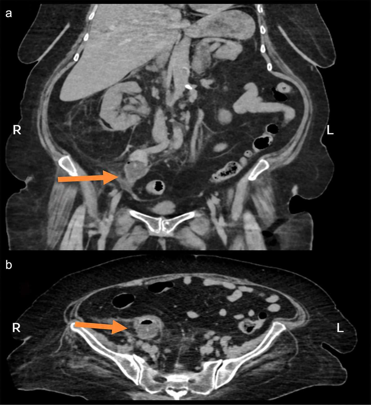 Click for large image | Figure 1. Preoperative computed tomography (CT) findings. (a) Coronal non-contrast CT image demonstrating the fully inflated Foley catheter balloon within the right ureter immediately below the iliac vessel crossing. (b) Axial non-contrast CT image confirming intraluminal balloon position within the right ureter. |
Under spinal anesthesia in the lithotomy position, cystoscopy was performed and the right ureteral orifice was entered. Ureteroscopy demonstrated extensive clot burden and a full-thickness ureteral wall defect located immediately distal to the iliac vessel crossing; the defect was estimated intraoperatively to be approximately 2.5 cm in length. Contrast injection showed complete extravasation, and a guidewire could not initially be advanced into the renal pelvis (Fig. 2). The intact lateral ureteral wall was carefully followed to identify the proximal true lumen, allowing for re-entry and access to the renal pelvis. Intrarenal contrast injection confirmed correct access.
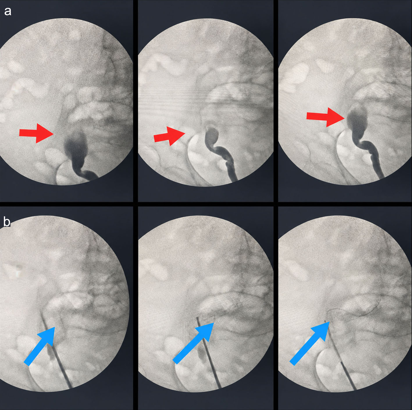 Click for large image | Figure 2. Intraoperative fluoroscopic findings. (a) Fluoroscopic image demonstrating contrast extravasation from the right ureter, consistent with full-thickness perforation. (b) Misguided guidewire advancement secondary to the ureteral wall defect. |
The decision to place two 6 Fr double-J stents was made intraoperatively. The relatively wide ureteral lumen and extensive full-thickness defect raised concern that a single stent might provide less stable drainage or migrate. Dual stenting was therefore chosen with the theoretical aims of increasing luminal stability, maintaining drainage if one stent became obstructed or displaced, providing dependable decompression across the injured segment, and preserving ureteral patency during healing (Fig. 3). These were intraoperative considerations rather than proven advantages over a single stent. Percutaneous nephrostomy was not selected initially because renal collecting-system dilatation was minimal and successful true-lumen access allowed for retrograde internal drainage. If retrograde stenting had failed, percutaneous nephrostomy was planned in consultation with interventional radiology. A new bladder catheter was placed at the end of the procedure.
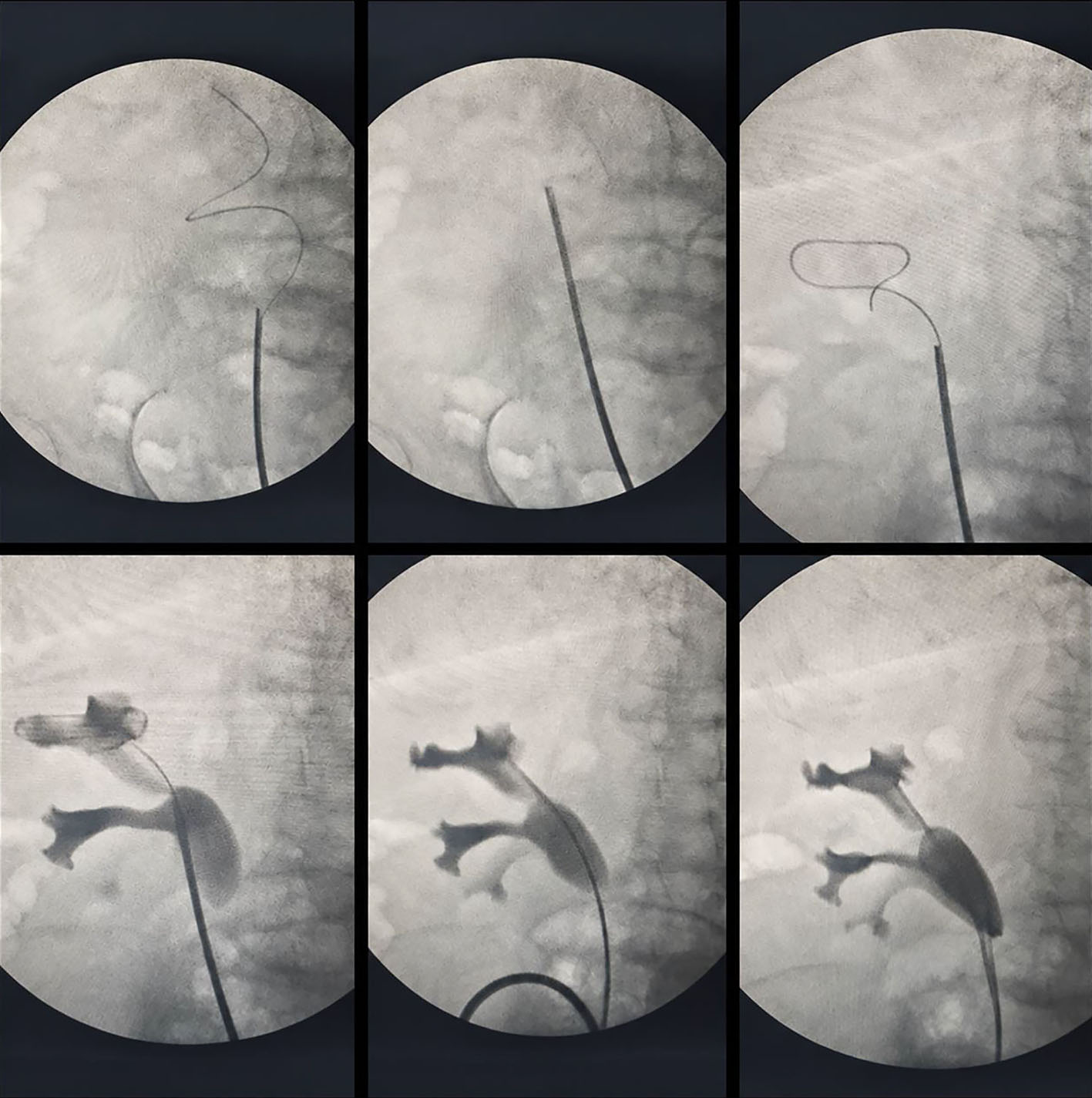 Click for large image | Figure 3. Intraoperative ureteroscopic and fluoroscopic findings. During ureterorenoscopy, the intact lateral ureteral wall was carefully followed to re-enter the true lumen and obtain renal pelvic access beyond the proximal margin of the perforation. Intrarenal localization was confirmed by contrast injection, and two 6 Fr double-J stents were placed for internal drainage and stabilization. |
Postoperative course
Empirical intravenous meropenem was initiated in consultation with infectious diseases and was continued under serial infectious diseases review for a total of 14 days. Dosing was adjusted to renal function; once serum creatinine decreased below 1 mg/dL, 1 g intravenously every 8 h was recommended. Two urine cultures obtained at presentation were reported as bacillary growth, but no species-level identification or antibiogram was available for the urine isolates. One blood culture grew Klebsiella pneumoniae ssp. pneumoniae. Susceptibility testing of the blood isolate showed resistance to meropenem and susceptibility to imipenem. Despite the in vitro meropenem resistance, meropenem was continued under serial infectious diseases consultation after successful urinary drainage and source control; no documented antimicrobial switch was identified in the available records. Follow-up blood and urine cultures obtained on treatment day 7 showed no growth.
On postoperative day 1, CRP increased to 355 mg/L, leukocyte count decreased to 19,000/µL, and serum creatinine improved to 1.7 mg/dL. By postoperative day 2, CRP had declined to 198 mg/L and leukocyte count to 13,000/µL. Gross hematuria resolved by postoperative day 3. Clinical and laboratory improvement continued during treatment, and by day 14 both the leukocyte count and CRP had normalized.
On postoperative day 2, triphasic CT at the level of the previous ureteral injury demonstrated regression of periureteral inflammatory changes and no evidence of urinary extravasation or urinoma. A plain kidney-ureter-bladder (KUB) radiograph confirmed appropriate in situ positioning of both right-sided double-J stents (Fig. 4).
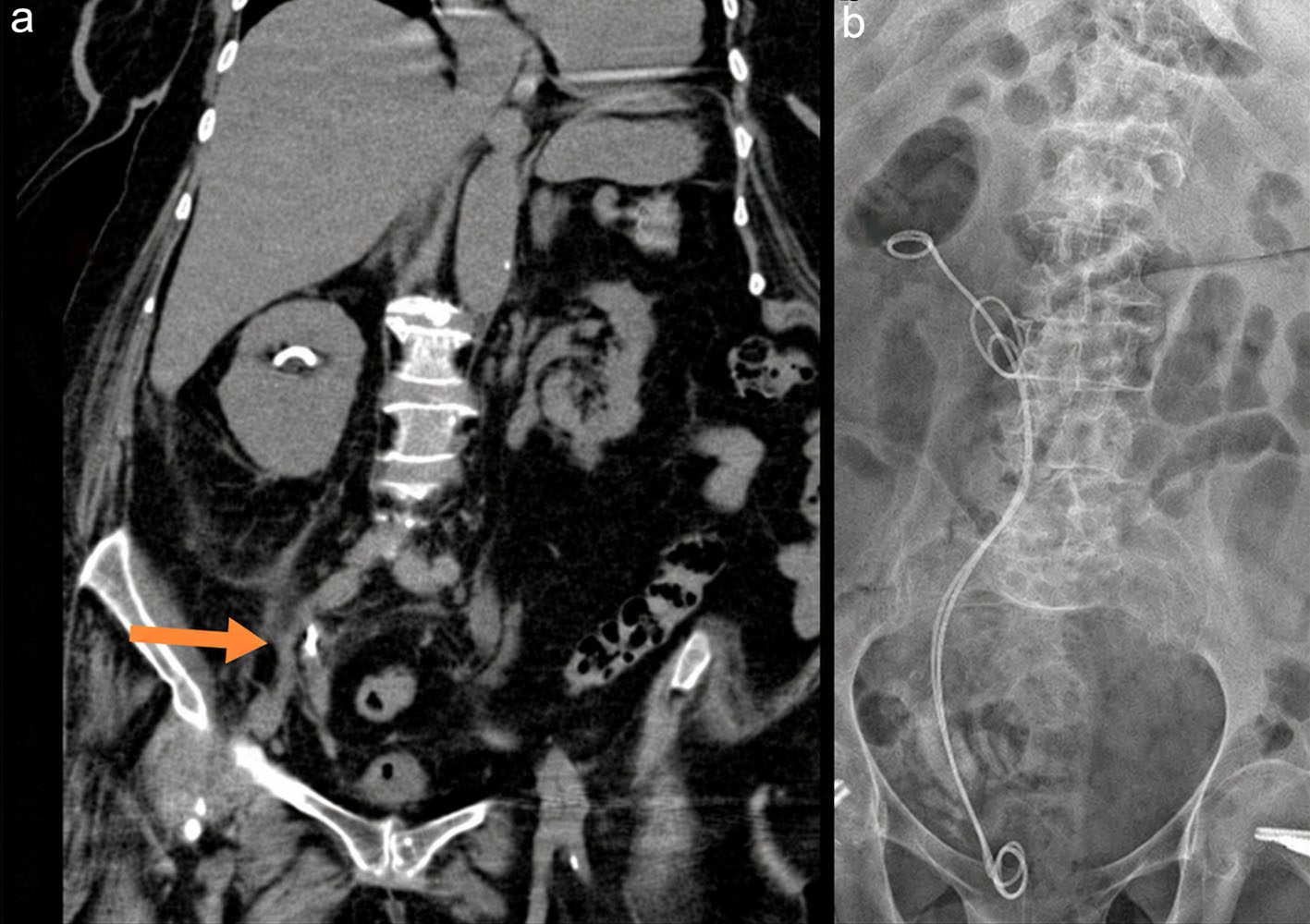 Click for large image | Figure 4. Early postoperative imaging findings. (a) Coronal image from triphasic computed tomography obtained on postoperative day 2 at the level of the previous ureteral injury (arrow), demonstrating regression of periureteral inflammatory changes and no evidence of urinary extravasation or urinoma. (b) Plain kidney-ureter-bladder radiograph demonstrating appropriate in situ positioning of both right-sided double-J stents. |
At 6 weeks, retrograde pyelography performed before planned stent removal demonstrated restoration of ureteral continuity without contrast extravasation. Delayed-phase contrast-enhanced CT (excretory phase) likewise showed no urinary leakage or urinoma (Fig. 5). Both double-J stents were then removed. The patient did not return for subsequent long-term follow-up; therefore, no 3- or 6-month post-removal ultrasound or CT was available to assess delayed ureteral stricture, hydronephrosis, or longer-term renal function; approximately 6 months had elapsed since discharge without a recorded follow-up visit.
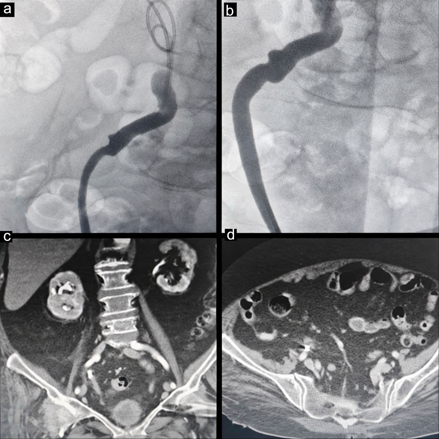 Click for large image | Figure 5. Retrograde pyelography at 6 weeks. (a, b) Retrograde pyelography showing restoration of ureteral continuity without contrast extravasation. (c, d) Delayed-phase contrast-enhanced CT images (excretory phase) demonstrating no evidence of urinary leakage or urinoma. CT: computed tomography. |
| Discussion | ▴Top |
Inadvertent ureteral cannulation by a urinary catheter remains rare, but a 2024 literature review of 48 reported patients found that 67% were female, 67% had long-term catheterization, and neurologic conditions were common; ureteral rupture occurred in a minority but clinically important proportion of cases [4]. The present patient had several converging risk factors: female sex, chronic catheterization, a clinically diagnosed neurogenic bladder, a cystoscopically documented bladder capacity below 100 mL, advanced dementia, and limited mobility. VUR has been proposed as a predisposing mechanism in the literature, but it was not demonstrated in this patient because no voiding cystourethrography had been performed.
Prevention depends on recognizing abnormal catheter behavior before balloon inflation. In patients with chronic catheters or altered bladder anatomy, absent or poor urine return, unexpected resistance, flank or abdominal pain, inability to irrigate freely, or poor return during irrigation should prompt concern for malposition [2, 5–7]. When ultrasonography is not immediately available, these bedside findings remain important practical checks; balloon inflation should be deferred if placement is uncertain, and catheter position should be confirmed by the most readily available appropriate imaging or endoscopic assessment.
The patient was hemodynamically stable at presentation, but fever, tachypnea, marked leukocytosis, elevated CRP, AKI, and gross hematuria indicated a clinically significant infected ureteral injury requiring prompt source control. Non-contrast CT was sufficient to demonstrate the Foley balloon within the ureter. Additional preoperative contrast imaging was not pursued because it was unlikely to change the immediate need for intervention and would have delayed drainage in the setting of AKI. After drainage and improvement in renal function, postoperative triphasic CT was used to document resolution of extravasation and regression of periureteral inflammation. More broadly, rapid recognition of urinary extravasation and timely decompression are central principles in acute urinary tract rupture [8, 9].
Management of iatrogenic ureteral injury is individualized according to injury continuity, location, timing, and the ability to establish drainage. Current European Association of Urology (EAU) guidance recognizes ureteral stenting and nephrostomy as urinary diversion options for selected ureteral injuries, while reconstructive repair is required when injury characteristics or failure of diversion make this necessary [8]. Hale et al described a mid-ureteral rupture managed with ureteroureteral anastomosis [10]. Modi et al reported unsuccessful retrograde stenting followed by antegrade stenting and nephrostomy [11]. In contrast, Locherbach et al and Doherty et al reported successful short-term healing after a single ureteral stent, with the latter report emphasizing continued surveillance for delayed narrowing [12, 13].
In the present case, the ureter was not completely transected; an intact lateral wall provided a visual landmark for true-lumen re-entry, and renal pelvic access was achieved retrogradely. Collecting-system dilatation was minimal. Once adequate internal drainage could be established, nephrostomy was not considered necessary as the initial approach, although it remained the planned rescue strategy if retrograde stenting failed. Similarly, immediate reconstructive surgery was not pursued because the injured segment could be bridged and drained endoscopically.
The choice of two simultaneous 6 Fr stents was based on intraoperative anatomy and surgeon judgment rather than evidence that dual stenting is superior. In a relatively wide ureter with an approximately 2.5-cm full-thickness defect, dual stents were hypothesized to provide more stable internal splinting, reduce the practical consequences of migration or obstruction of one stent, and maintain decompression and patency across the injured segment. These mechanisms were not directly measured in this patient and should be interpreted as the rationale for the technique, not as demonstrated physiological benefits. In the reviewed literature on Foley-associated ureteral injury, we did not identify a prior report describing two simultaneous double-J stents for a balloon-induced full-thickness perforation; previously reported severe cases were managed with reconstruction, nephrostomy/antegrade stenting, or a single ureteral stent [4, 10–13].
Short-term healing was demonstrated by the absence of extravasation on postoperative imaging and by retrograde pyelography at 6 weeks. However, delayed ureteral stricture is a clinically important endpoint after a full-thickness injury. The absence of 3- or 6-month imaging is therefore a major limitation of this report, and long-term ureteral patency cannot be inferred from the 6-week result. Future similar cases should undergo imaging surveillance after stent removal, for example, at 3 and/or 6 months, to assess hydronephrosis, ureteral narrowing, and renal function.
Conclusion
Inadvertent ureteral placement and balloon inflation of a Foley catheter can cause a severe full-thickness ureteral injury, particularly in patients with chronic catheterization and markedly altered bladder anatomy. In this selected patient, endoscopic true-lumen re-entry and dual double-J stenting achieved short-term urinary drainage and healing without immediate nephrostomy or reconstruction. Dual stenting should be regarded as a potential technical option rather than a proven superior strategy; choice of drainage must remain individualized, with nephrostomy or reconstruction available when retrograde drainage is unsuccessful or inadequate. Longer-term imaging follow-up is required to assess delayed stricture.
Acknowledgments
None to declare.
Financial Disclosure
No external funding was received for this study.
Conflict of Interest
The author declares no conflict of interest.
Informed Consent
Written informed consent for the publication of this case report and accompanying images were obtained from the patient’s legal representative.
Author Contributions
Ibrahim Erol was solely responsible for the conception of the case report, data curation, investigation, writing – original draft, and writing–review and editing.
Data Availability
Data supporting the findings of this case report are not publicly available to protect patient privacy, but may be available from the corresponding author upon reasonable request, subject to ethical and institutional requirements.
AI Use Declaration
During manuscript preparation, ChatGPT (OpenAI) was used for language editing and organizational assistance. The author reviewed and revised all content and takes full responsibility for the accuracy, integrity, and originality of the submitted manuscript.
Abbreviations
AKI: acute kidney injury; EAU: European Association of Urology; CRP: C-reactive protein; CT: computed tomography; KUB: kidney-ureter-bladder; VUR: vesicoureteral reflux
| References | ▴Top |
- Baker KS, Dane B, Edelstein Y, Malhotra A, Gould E. Ureteral rupture from aberrant Foley catheter placement: a case report. J Radiol Case Rep. 2013;7(1):33-40.
doi pubmed - Al-Zubi M, Alheyassat MMF, Alhasan M, Al-Qudah MHM, Bani-Hani M. Unintended Foley's catheter placement into the ureter: a case report. Int J Surg Case Rep. 2022;100:107750.
doi pubmed - Cho SK, Kim MS, Chung HS, Hwang EC, Jung SI, Kwon D, Park K. Transurethral Foley catheter misplacement into the upper urinary tract in a patient with a history of lung cancer and chemotherapy: a case report and considerations to keep in mind. Transl Androl Urol. 2021;10(3):1347-1351.
doi pubmed - Li JJ, Au CF. Inappropriate placement of urinary catheters into the ureter: a case report and literature review. Medicine (Baltimore). 2024;103(15):e37623.
doi pubmed - Anderson BW, Greenlund AC. Ureteral cannulation as a complication of urethral catheterization. Korean J Urol. 2014;55(11):768-771.
doi pubmed - Luo R, Lee SL, Ng FC, Koh LT. Inadvertent placement of a urinary catheter into the ureter: a report of 3 cases and review of the literature. Asian J Urol. 2017;4(4):256-261.
doi pubmed - Ishikawa T, Araki M, Hirata T, Watanabe M, Ebara S, Watanabe T, Nasu Y, et al. A rare complication: misdirection of an indwelling urethral catheter into the ureter. Acta Med Okayama. 2014;68(1):47-51.
doi pubmed - European Association of Urology (EAU). EAU Guidelines on Urological Trauma 2026. Arnhem: EAU Guidelines Office; 2026.
- Yorgun M, Duran AM, Aktas RS, Tas O. Spontaneous forniceal rupture in the emergency department. J Emerg Med Case Rep. 2025;16(4):130-133.
doi - Hale N, Baugh D, Womack G. Mid-ureteral rupture: a rare complication of urethral catheterization. Urology. 2012;80(5):e65-66.
doi pubmed - Modi PK, Salmasi AH, Perlmutter MA. Inadvertent foley catheterization of the ureter. Can J Urol. 2014;21(3):7326-7329.
pubmed - Locherbach F, Grimsehl P, Sauer A, Wyler S, Kwiatkowski M. Ureteral rupture caused by accidental intubation of the ureter with a foley-catheter during ureterorenoscopy. Case Rep Urol. 2018;2018:9738265.
doi pubmed - Doherty KSG, Melman EM, Kamphuis GM. Case of the Month from the University of Amsterdam UMC, the Netherlands: ureteric injury by a Foley catheter, an unusual complication in pregnancy. BJU Int. 2021;128(5):551-554.
doi pubmed
This article is distributed under the terms of the Creative Commons Attribution 4.0 International License (CC BY 4.0), which permits unrestricted use, distribution, and reproduction in any medium, including commercial use, provided the original work is properly cited.
World Journal of Nephrology and Urology is published by Elmer Press Inc.