Figures
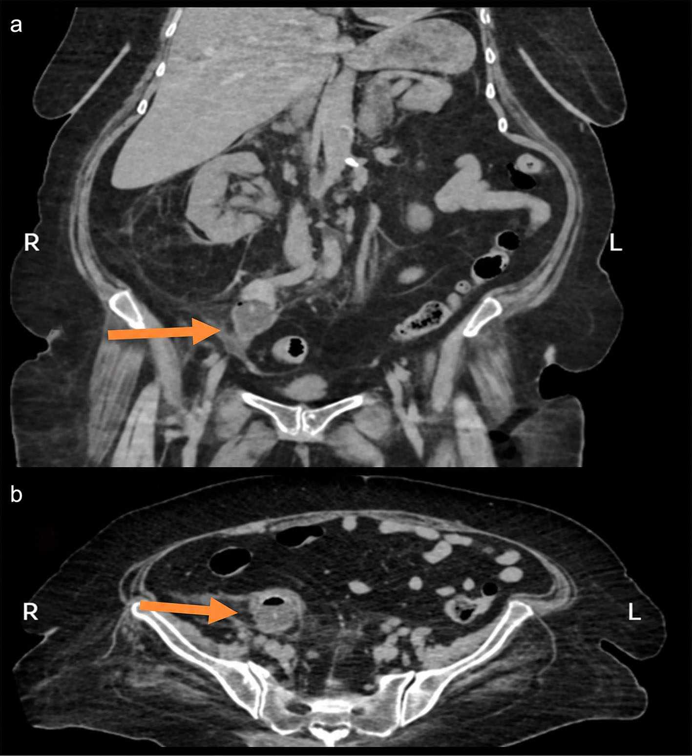
↓ Figure 1. Preoperative computed tomography (CT) findings. (a) Coronal non-contrast CT image demonstrating the fully inflated Foley catheter balloon within the right ureter immediately below the iliac vessel crossing. (b) Axial non-contrast CT image confirming intraluminal balloon position within the right ureter.
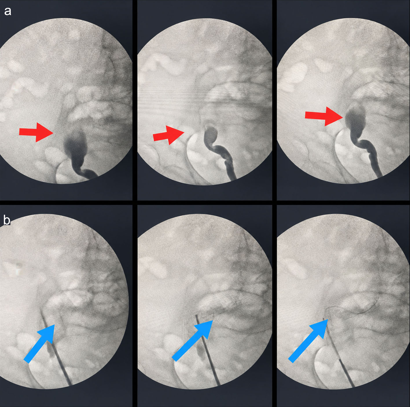
↓ Figure 2. Intraoperative fluoroscopic findings. (a) Fluoroscopic image demonstrating contrast extravasation from the right ureter, consistent with full-thickness perforation. (b) Misguided guidewire advancement secondary to the ureteral wall defect.
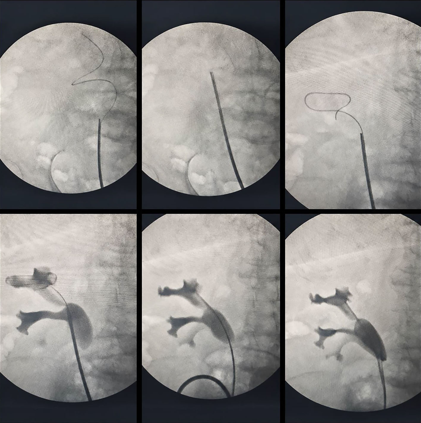
↓ Figure 3. Intraoperative ureteroscopic and fluoroscopic findings. During ureterorenoscopy, the intact lateral ureteral wall was carefully followed to re-enter the true lumen and obtain renal pelvic access beyond the proximal margin of the perforation. Intrarenal localization was confirmed by contrast injection, and two 6 Fr double-J stents were placed for internal drainage and stabilization.
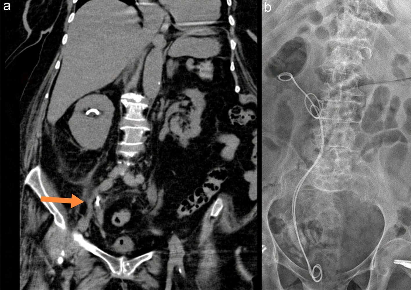
↓ Figure 4. Early postoperative imaging findings. (a) Coronal image from triphasic computed tomography obtained on postoperative day 2 at the level of the previous ureteral injury (arrow), demonstrating regression of periureteral inflammatory changes and no evidence of urinary extravasation or urinoma. (b) Plain kidney-ureter-bladder radiograph demonstrating appropriate in situ positioning of both right-sided double-J stents.
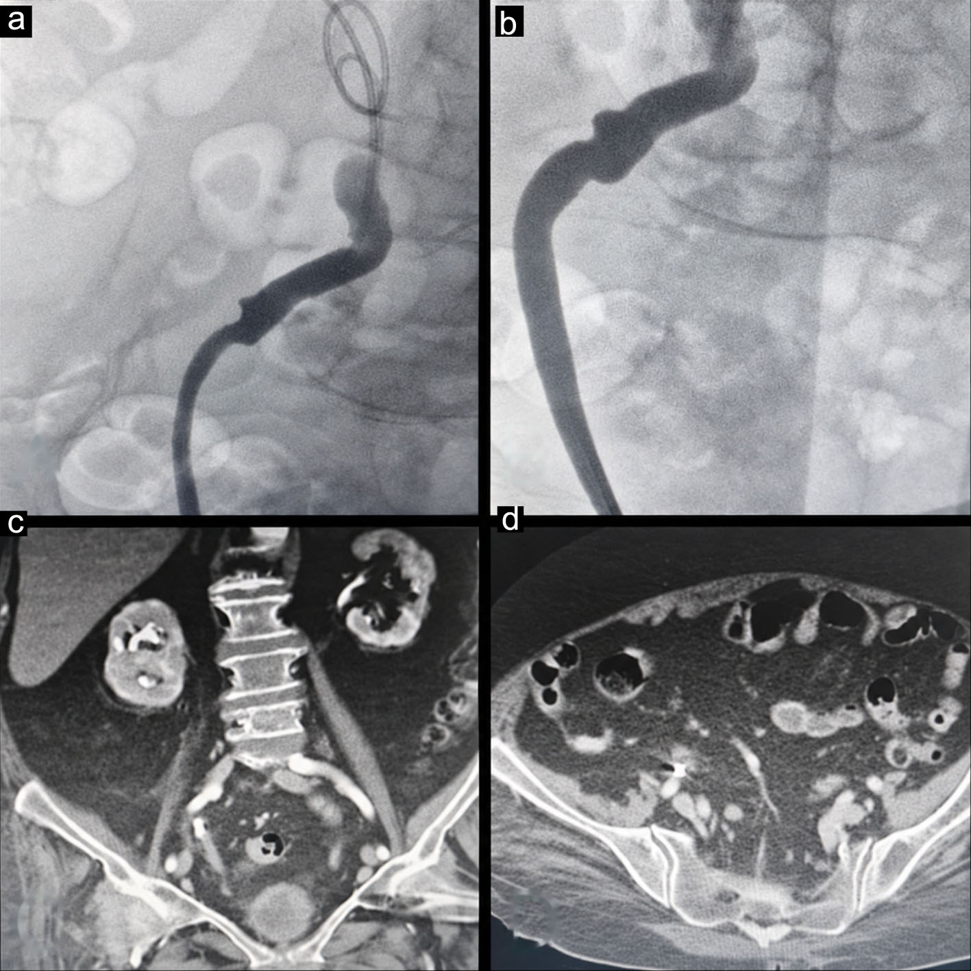
↓ Figure 5. Retrograde pyelography at 6 weeks. (a, b) Retrograde pyelography showing restoration of ureteral continuity without contrast extravasation. (c, d) Delayed-phase contrast-enhanced CT images (excretory phase) demonstrating no evidence of urinary leakage or urinoma. CT: computed tomography.