| World Journal of Nephrology and Urology, ISSN 1927-1239 print, 1927-1247 online, Open Access |
| Article copyright, the authors; Journal compilation copyright, World J Nephrol Urol and Elmer Press Inc |
| Journal website https://wjnu.elmerpub.com |
Case Report
Volume 15, Number 4, November 2026, pages 125-130

Inguinoscrotal Herniation of the Urinary Bladder Presenting With Obstructive Uropathy: A Rare but Important Diagnostic Consideration
Apoorva Tripathia, d  , Beril Isa, Octavio Jose Del Realb , Sarp Keskina, c
aDepartment of Urology, Oxford University Hospitals NHS Foundation Trust, Oxford, UK
bDepartment of Urology, Hospital Felix Bulnes Cerda, Santiago, Chile
cDepartment of Urology, Koc University School of Medicine, Istanbul, Turkey
dCorresponding Author: Apoorva Tripathi, Department of Urology, Oxford University Hospitals NHS Foundation Trust, Oxford, UK
Manuscript submitted June 22, 2026, accepted October 5, 2026, published online November 18, 2026
Short title: Inguinoscrotal Bladder Hernia With Uropathy
doi: https://doi.org/10.14740/wjnu1062
| Abstract | ▴Top |
Bladder involvement within an inguinal hernia is rare, occurring in fewer than 4% of cases, and is seldom recognized before operation. When the herniated segment is large, it can produce serious morbidity, including obstructive uropathy and deteriorating renal function, so timely recognition is important both to avoid intraoperative bladder injury and to reverse renal compromise. We report a 71-year-old man with a longstanding right scrotal swelling, nocturnal urinary leakage, an obligatory seated voiding posture, and rapidly worsening renal function. Ultrasound demonstrated a large pelvic fluid collection extending into the scrotum, and contrast-enhanced computed tomography (CT) confirmed a right inguinoscrotal hernia containing most of the urinary bladder, with bilateral ureteric compression at the vesicoureteric junctions and consequent hydronephrosis. The estimated glomerular filtration rate (eGFR) fell from 78 to 48 mL/min/1.73 m2, with a corresponding rise in serum creatinine, indicating acute kidney injury. Urethral catheterization drained a large residual volume, produced immediate decompression and a striking reduction in scrotal swelling, and served as a useful early diagnostic sign. The patient underwent urgent open right inguinal hernia repair; operative findings showed coexisting direct and indirect components, with the bladder lying within the direct defect. The bladder was reduced without resection, the posterior wall reconstructed, and a polypropylene mesh placed, with intraoperative cystography confirming bladder integrity. Recovery was uneventful and renal function had normalized by 8 months with no recurrence. This case is instructive because it links a frequently overlooked anatomical problem to a reversible cause of renal failure, and it reinforces the value of ultrasound as the first-line investigation that raised the initial suspicion of bladder herniation and of CT in delineating hernia contents and protecting the bladder during repair. The principal learning points are to suspect bladder herniation in older, obese men with large inguinal hernias and atypical urinary symptoms such as positional voiding, to obtain CT when urinary features accompany a groin hernia, and to recognize that prompt decompression and repair can restore both urinary and renal function.
Keywords: Inguinoscrotal bladder hernia; Scrotal cystocele; Obstructive uropathy; Acute kidney injury; Bilateral hydronephrosis; Inguinal hernia repair; Computed tomography; Mesh hernioplasty
| Introduction | ▴Top |
Groin hernias rank among the most common abdominal wall defects in surgical practice and occur disproportionately in older men [1, 2]. A hernial sac most often contains omentum or small bowel; the urinary bladder is an unusual occupant, described in under 4% of inguinal hernias [1, 2]. Known also as scrotal cystocele or inguinoscrotal bladder hernia, it develops chiefly in older men in whom long-standing bladder outlet obstruction, obesity, and gradual attenuation of the abdominal wall together draw the bladder into the defect over months to years [1, 3, 4]. Because it is seldom suspected before operation, an extensively herniated bladder can escape notice until appreciable morbidity has appeared, ranging from voiding dysfunction to, in severe cases, obstructive uropathy with renal impairment [1, 5].
We present a 71-year-old man with a large right inguinoscrotal hernia containing the majority of the urinary bladder, complicated by bilateral hydronephrosis and acute kidney injury [4, 6]. The case is reported to illustrate two points of practical importance. First, a high index of suspicion is required in older patients with large or longstanding inguinal hernias when accompanied by atypical urinary symptoms, since the characteristic features are easily dismissed as benign prostatic disease or age-related change [1, 7]. Second, cross-sectional imaging, in particular computed tomography (CT), is central to preoperative planning because it defines the hernia contents and ureteric involvement, allows the surgeon to anticipate abnormal anatomy, and thereby reduces the risk of inadvertent bladder injury during repair [1, 8]. Ultrasound is frequently the first imaging study obtained for scrotal swelling and, as in the present case, may be the investigation that first raises the possibility of bladder herniation, prompting confirmatory cross-sectional imaging.
| Case Report | ▴Top |
Investigations (patient information and clinical findings)
A 71-year-old man presented with a longstanding right scrotal swelling. He reported nocturnal urinary leakage and was able to void only while seated. He described urinary urgency and frequency but denied dysuria or hematuria at presentation. His past medical history included hypertension, previous myocardial infarction, and obesity, with a body mass index of 36.3 kg/m2. He smoked two cigarettes daily.
There was no relevant family history, and no relevant genetic or psychosocial factors were identified. He had no history of prior inguinal hernia repair or other relevant urological intervention.
On examination, there was a massively enlarged, non-tender right scrotum that was continuous with the inguinal canal. Initial laboratory testing showed a mildly reduced estimated glomerular filtration rate (eGFR) of 78 mL/min/1.73 m2 with a serum creatinine of 86 µmol/L. Prostate-specific antigen (PSA) was unremarkable. The chronology of presentation, investigation, treatment, and recovery is summarized in Table 1.
 Click to view | Table 1. Timeline of Clinical Events |
Diagnosis
The diagnosis was reached through a combination of physical examination, laboratory testing, and imaging. Ultrasound of the right scrotum demonstrated a large fluid collection exceeding 800 mL that extended into the pelvis, raising the possibility of bladder herniation (Fig. 1). The ultrasound was directed at the scrotum and pelvis and did not include dedicated views of the kidneys; the hydronephrosis was therefore not identified on ultrasound and was first demonstrated on the subsequent cross-sectional imaging. Contrast-enhanced CT of the abdomen and pelvis then confirmed a large right inguinal hernia containing the majority of the bladder (Fig. 2), with compression of both distal ureters at the vesicoureteric junctions and resultant bilateral hydronephrosis.
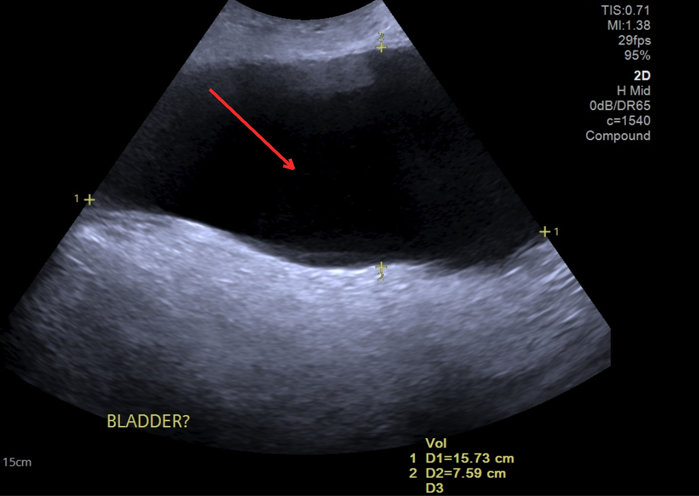 Click for large image | Figure 1. Transabdominal and scrotal ultrasound. Large anechoic, fluid-filled structure exceeding 800 mL extending from the pelvis into the scrotum (red arrow), initially reported as a query bladder and in keeping with the herniated urinary bladder. Ultrasound was the first-line study and raised the initial suspicion of bladder herniation, before cross-sectional imaging was obtained. |
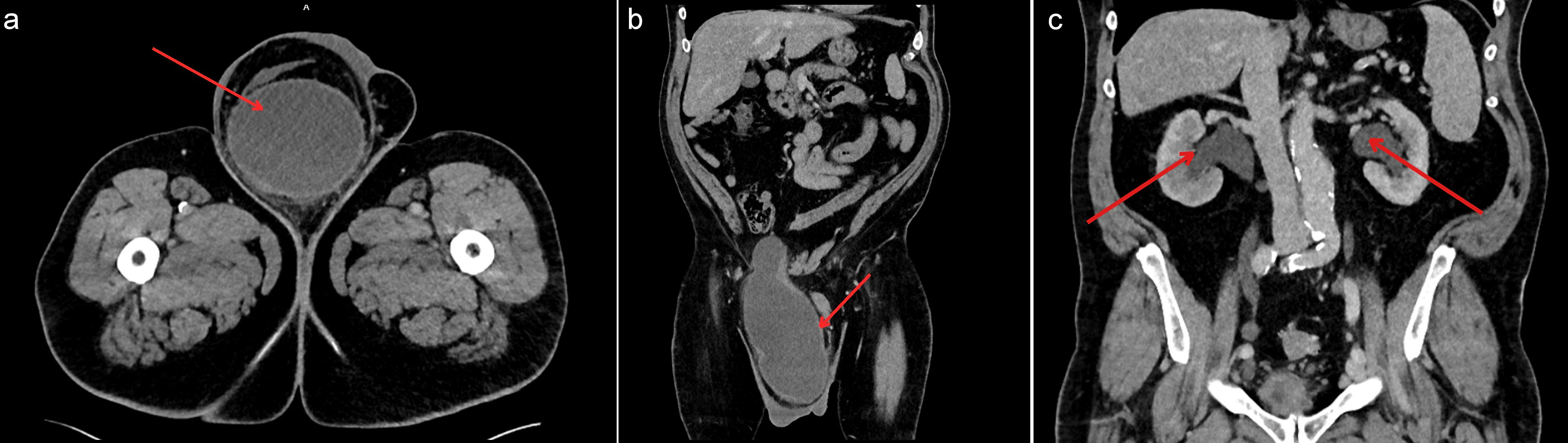 Click for large image | Figure 2. Contrast-enhanced CT of the abdomen and pelvis. (a) Axial image showing a large right inguinoscrotal hernia containing the majority of the urinary bladder. (b) Coronal image showing descent of the bladder from the pelvis into the right scrotum. (c) Coronal image at the level of the kidneys demonstrating bilateral hydronephrosis with dilatation of the renal pelves and proximal ureters (red arrows indicate the dilated pelvicalyceal systems). In panels (a) and (b), the red arrow indicates the herniated bladder. CT: computed tomography. |
The principal diagnostic challenge was that the presenting symptoms overlapped closely with common lower urinary tract complaints and could readily have been attributed to benign prostatic disease. The obligatory seated voiding and nocturnal leakage were the atypical features that prompted closer scrutiny. The bladder origin of the scrotal fluid collection was further supported when urethral catheterization produced immediate decompression and a dramatic reduction in scrotal swelling, which served as a valuable confirmatory sign. Catheterization also produced a substantial reduction in the herniated intrascrotal component itself. Because the hernia was composed largely of the fluid-filled bladder, draining the bladder decompressed and partially reduced the hernia; this is the expected response in a sliding bladder hernia and is itself a useful diagnostic clue, since a hydrocele or a bowel-containing hernia does not resolve with urinary catheterization.
Differential diagnosis
Other causes of a large scrotal swelling were considered, including hydrocele, an inguinoscrotal hernia containing bowel or omentum, and a scrotal abscess or other fluid collection. The combination of imaging appearances, the urinary symptom profile, and the response to catheterization distinguished bladder herniation from these alternatives.
Prognosis
The prognosis was judged favorable provided obstruction was relieved promptly, because obstructive uropathy of recent onset is frequently reversible when the obstructing cause is corrected without delay.
Treatment
Emergency urethral catheterization was performed first as a decompressive measure; it drained 500 to 600 mL of urine, produced immediate relief of the herniated component, and reduced upstream ureteric pressure. The patient then underwent urgent open right inguinal hernia repair. Operative findings revealed coexisting direct and indirect hernias, with the bladder lying within the direct component. The hernia was fully reducible. The bladder was dissected free and reduced into the pelvis without resection, the posterior wall was reconstructed, and a tension-free polypropylene mesh repair was performed. Intraoperative cystography confirmed bladder integrity. A pelvic drain was placed, and the procedure was completed without complication.
Follow-up and outcomes
Postoperative recovery was uneventful. Mild hematuria on the first postoperative day was self-limiting. The urethral catheter was retained for 5 days to allow the manipulated bladder wall to heal and was then removed without difficulty. The wound remained clean and the patient was discharged on the second postoperative day.
At 2 weeks, flexible cystoscopy was undertaken to exclude intravesical pathology in the context of the postoperative hematuria and the anatomical distortion produced by prolonged herniation; this was unremarkable, and the patient underwent a successful trial without catheter and voided satisfactorily. By 8 months, renal function had fully normalized, with an eGFR greater than 90 mL/min/1.73 m2, and there were no further urinary complications and no hernia recurrence. Intervention adherence and tolerability were assessed at routine clinical follow-up, and no adverse or unanticipated events occurred.
| Discussion | ▴Top |
Inguinal bladder herniation is infrequent yet carries real clinical weight. Pooled data from a systematic review, covering 64 patients reported across 51 articles, place its frequency below 4% of inguinal hernias and confirm a bias toward older, obese men with bladder outlet obstruction [1–3]. The present patient embodied these well-documented risk factors: advanced age, obesity, lower urinary tract symptoms, and a longstanding scrotal swelling [1, 4, 5]. His obligatory seated voiding posture and nocturnal leakage are characteristic adaptive behaviors that help to distinguish bladder herniation from benign prostatic disease [3, 6]. Such features are easily dismissed as age-related change, and their overlap with ordinary lower urinary tract symptoms explains why the diagnosis is so often delayed. A single atypical feature, such as positional voiding, should prompt closer scrutiny rather than reassurance.
Although bladder involvement complicates fewer than 4% of inguinal hernias, it is probably underrecognized rather than genuinely rare. It should be acknowledged that, once a patient with acute kidney injury undergoes dedicated renal ultrasound, the associated hydronephrosis is usually readily detected; the difficulty in bladder herniation therefore lies less in demonstrating upper tract dilatation than in recognizing the herniated bladder as its underlying cause, particularly when the initial ultrasound is directed at the scrotum and does not include the kidneys, as occurred in the present case. A growing literature documents its association with upper urinary tract obstruction. In the systematic review by Branchu et al, a substantial proportion of reported bladder hernias were accompanied by hydronephrosis or impaired renal function, yet preoperative recognition remained low [1]. Davis et al described complete inguinoscrotal bladder herniation with ureteric involvement and obstructive change [4]. Kraft et al, in a series with an accompanying review, similarly reported cases presenting with obstructive uropathy and renal impairment that improved after reduction and repair [9]. Reports of massive or giant hernias, including those complicated by intravesical stones or managed by laparoscopic and totally extraperitoneal techniques, have likewise noted secondary hydronephrosis and large post-void residual volumes [3, 5, 6]. Taken together, these reports indicate that obstructive uropathy is a recognized, if under-reported, complication of large bladder hernias, and that renal function commonly recovers when the obstruction is relieved without delay. The present case adds to this literature by documenting the complete biochemical and radiological trajectory from acute kidney injury through to full renal recovery.
The mechanism reflects the partly extraperitoneal position of the bladder, which can be drawn into a hernial defect by traction from a peritoneal sac or pushed through a weakened abdominal wall as intra-abdominal pressure rises. Chronic obstruction, obesity, and pelvic floor laxity generate a sustained pressure gradient that gradually displaces the bladder over months to years [1, 3, 4]. Depending on how the bladder relates to the peritoneal sac, these hernias are grouped as paraperitoneal, intraperitoneal, or extraperitoneal, a scheme that matters surgically because it dictates the dissection plane and the likelihood of accidental cystotomy [7]. The coexistence of direct and indirect components in this patient illustrates how mixed hernias complicate the anatomy and demand careful intraoperative identification of bladder tissue before any sac is divided.
A particularly important feature of this case was the extent of bladder descent, which was sufficient to cause bilateral hydronephrosis and rapid renal deterioration. As the bladder herniates, it draws the trigone and ureterovesical junctions inferiorly, kinking the distal ureters and producing functional obstruction [4, 7, 9]. For much of the natural history, this process is slow and compensated: the upper urinary tracts accommodate gradually rising pressures, and renal function is preserved. The development of acute kidney injury on this background of a longstanding, previously stable hernia is one of the most instructive aspects of the case, and it reflects the crossing of a mechanical threshold. As the herniated bladder volume increases, the trigone and both ureterovesical junctions become fixed in a progressively more angulated position within the inguinal canal, while incomplete emptying and a rising post-void residual further distend the intrascrotal bladder and raise intravesical and intraureteric pressure. Beyond a critical point, the bilateral vesicoureteric obstruction can no longer be compensated, and an episode of acute-on-chronic retention converts chronic partial obstruction into acute bilateral hydronephrosis. In this patient, the combination of a very large, herniated volume, obligatory seated voiding with incomplete bladder emptying, and a rising residual is likely to have produced precisely such a tipping point, which explains why the obstruction became clinically significant at this particular time. The decline in eGFR from 78 to 48 mL/min/1.73 m2 demonstrates how an apparently chronic groin swelling can become an acute emergency. Immediate catheterization drained a large residual volume, decompressed the herniated component, and relieved upstream ureteric pressure, in keeping with established emergency principles, and the marked reduction in scrotal swelling that followed was itself a valuable diagnostic sign [2, 10].
The complete recovery of renal function by 8 months illustrates a reassuring principle: obstructive uropathy is frequently reversible when the obstruction is identified and relieved promptly [9, 11]. The duration and degree of obstruction are key determinants of renal recovery, which provides a powerful argument for early recognition [11]. Bladder herniation may remain asymptomatic for years and has historically been identified before operation in fewer than 10% of cases; an unsuspected bladder within a hernial sac is therefore at substantial risk of iatrogenic injury during dissection [1, 7].
Ultrasound was the pivotal first-line investigation in this case and deserves emphasis: it was the modality that initially raised the suspicion of bladder herniation by revealing a large anechoic, fluid-filled structure in continuity with the pelvis, which then prompted confirmatory cross-sectional imaging. Scrotal ultrasound is widely available, radiation-free, and inexpensive, and it can reliably distinguish a fluid-filled herniated bladder from a hydrocele or a bowel-containing hernia, particularly when real-time compression, color Doppler, and reassessment after catheter drainage are used. The observation that the intrascrotal fluid volume fell markedly once the bladder was catheterized provided further bedside confirmation of the bladder origin of the swelling. Its principal limitations are operator dependence and reduced precision in delineating the surgical anatomy. Contrast-enhanced CT is complementary and provides the clearest delineation of hernia contents, ureteric involvement, and associated complications such as hydronephrosis and diverticula, and it remains the most informative modality for preoperative planning [1, 7, 8]. Preoperative identification of bladder involvement markedly reduces intraoperative risk by alerting the surgeon to decompress the bladder and to anticipate the abnormal anatomy [12].
Open repair remains the preferred approach for large, complex hernias because it allows direct visualization, controlled reduction, and reliable mesh reinforcement [1, 5, 13]. Laparoscopic and totally extraperitoneal techniques have been used successfully for smaller hernias in selected patients [6, 14]. The choice of an open approach in this patient merits explanation. Laparoscopic and totally extraperitoneal repairs offer genuine advantages, including magnified direct visualization of the herniated bladder, accurate assessment of the hernia orifice, and clear delineation of the direct and indirect components. Several features of this case nonetheless favored open repair. The hernia was very large and contained the majority of the bladder, the defect was mixed with coexisting direct and indirect components, and the patient presented acutely with obstructive uropathy and acute kidney injury that required prompt decompression. An open approach permitted controlled dissection of the bladder from the sac, safe reduction without resection, direct reconstruction of the posterior wall, tension-free mesh placement, and immediate intraoperative cystography to confirm bladder integrity, while minimizing the risk of iatrogenic cystotomy in markedly distorted anatomy. It also avoided the pneumoperitoneum and the prolonged operative time that a complex laparoscopic reduction of a giant hernia would entail in a comorbid patient with recent renal compromise. For these reasons, and in keeping with the literature that favors open repair for large, complex, or acutely presenting bladder hernias [1, 5, 13], an open tension-free mesh repair was judged the safest and most definitive option. Bladder resection is rarely required and is reserved for cases involving necrosis, large diverticula, or nonviable tissue; simple reduction of a viable bladder followed by tension-free mesh repair, as here, is both appropriate and definitive, and intraoperative cystography to confirm bladder integrity is a prudent step whenever the bladder has been mobilized [1, 5, 15]. Postoperative outcomes are generally favorable: mild, self-limiting hematuria after bladder manipulation is well described and rarely requires intervention [1, 2]; catheter drainage for 3 to 5 days allows the bladder wall to heal and reduces tension on the repair [5]; and reported long-term recurrence rates after mesh repair are low, generally in the range of 0% to 5%, with good resolution of urinary symptoms [1, 3, 5, 16].
Strengths and limitations
The strengths of this report lie in the complete clinical, biochemical, radiological, operative, and follow-up data, which together document the full course from atypical presentation to renal recovery. As a single case, it cannot establish incidence or guide management algorithms, and the conclusions drawn are necessarily based on this experience together with the existing literature.
In summary, this case reinforces that mechanical factors should be considered in older men with atypical lower urinary tract symptoms, since a proportion presumed to have benign prostatic hyperplasia may in fact harbor a bladder herniation that will not respond to prostate-directed therapy [17]. The rationale for our conclusions rests on the clear temporal link between herniation, ureteric obstruction, and renal decline, and on the prompt and complete reversal of that decline once the bladder was decompressed and the hernia repaired.
Conclusions
Inguinoscrotal herniation of the bladder, although rare, can convert a chronic groin swelling into an acute renal emergency. Clinicians should keep it in mind whenever an older or obese man presents with a large inguinal hernia together with atypical voiding symptoms or otherwise unexplained renal decline. Cross-sectional imaging, ideally contrast-enhanced CT, defines the anatomy, protects the bladder at operation, and supports a planned repair. As this case shows, timely decompression followed by tension-free mesh repair can fully reverse obstructive uropathy and return renal function to normal, and wider recognition of the condition should improve both diagnostic accuracy and operative safety.
Learning points
Bladder involvement should be suspected in older, obese men with large inguinoscrotal hernias and atypical urinary symptoms, particularly positional or seated voiding, nocturnal leakage, or unexplained renal deterioration, because these features are easily mistaken for benign prostatic disease, and the diagnosis is frequently delayed. Contrast-enhanced CT is the key investigation when urinary symptoms accompany a groin hernia, as it defines the hernia contents and ureteric involvement, guides surgical planning, and helps prevent intraoperative bladder injury. A striking reduction in scrotal swelling after urethral catheterization is a useful bedside diagnostic clue and an immediate decompressive measure. Prompt surgical repair with preoperative catheter decompression can reverse obstructive uropathy and restore both urinary and renal function, and because the duration of obstruction largely determines renal recovery, early recognition offers the best chance of a complete return of function.
Acknowledgments
None to declare.
Financial Disclosure
This research received no specific grant from any funding agency in the public, commercial, or not-for-profit sectors.
Conflict of Interest
The authors declare that they have no conflict of interest.
Informed Consent
Written informed consent for case publication was obtained from the patient, including consent for clinical images and imaging studies.
Author Contributions
Apoorva Tripathi conceived the report, collected and interpreted the clinical and imaging data, performed the literature review, and drafted the manuscript. Beril Is contributed to data acquisition, manuscript drafting, and revision. Octavio Jose Del Real contributed to the literature review and critical revision of the manuscript. Sarp Keskin was involved in the clinical and surgical management of the patient, supervised the work, and critically revised the manuscript for important intellectual content. All authors read and approved the final version of the manuscript.
Data Availability
The data supporting the findings of this case report are available from the corresponding author on reasonable request.
AI Use Declaration
An AI-based large language model tool was used to assist with language editing and formatting of the manuscript. It was not used to generate clinical data, analyses, interpretations, or conclusions. The authors reviewed and verified all content and accept full responsibility for the manuscript.
Abbreviations
AKI: acute kidney injury; BMI: body mass index; CT: computed tomography; eGFR: estimated glomerular filtration rate; PSA: prostate-specific antigen
| References | ▴Top |
- Branchu B, Renard Y, Larre S, Leon P. Diagnosis and treatment of inguinal hernia of the bladder: a systematic review of the past 10 years. Turk J Urol. 2018;44(5):384-388.
doi pubmed - Mohammadi A, Golmohammadi P, Pakdel A, Aghamir SMK. Laparoscopic management of a recurrent, inguinal hernia containing urinary bladder: a case report and literature review. Case Rep Med. 2025;2025:2076137.
doi pubmed - Sripathi S, Farooqi N, Kamel MK, Baroody R, Alashari A. Inguinoscrotal Hernia of the Urinary Bladder. Cureus. 2024;16(3):e56636.
doi pubmed - Davis DA, Glinski RW, Jones MC. A rare case of complete inguinoscrotal bladder herniation with ureteric involvement: assessing diagnostic challenges and complex surgical management. Cureus. 2024;16(2):e55130.
doi pubmed - Kutsal C, Baloglu IH, Cekmece AE, Ektiren M, Demir U. Bladder stones in a herniated bladder: A rare case report and literature review. Int J Surg Case Rep. 2022;93:106877.
doi pubmed - Chang HH, Wen SC. Extraperitoneal laparoscopic repair of huge inguinoscrotal bladder hernia: A case report and literature review. Urol Case Rep. 2021;38:101735.
doi pubmed - Elkbuli A, Narvel RI, McKenney M, Boneva D. Inguinal bladder hernia: A case report and literature review. Int J Surg Case Rep. 2019;58:208-211.
doi pubmed - Cabarrus MC, Yeh BM, Phelps AS, Ou JJ, Behr SC. From Inguinal Hernias to Spermatic Cord Lipomas: Pearls, Pitfalls, and Mimics of Abdominal and Pelvic Hernias. Radiographics. 2017;37(7):2063-2082.
doi pubmed - Kraft KH, Sweeney S, Fink AS, Ritenour CW, Issa MM. Inguinoscrotal bladder hernias: report of a series and review of the literature. Can Urol Assoc J. 2008;2(6):619-623.
doi pubmed - Thotakura R, Anjum F. Hydronephrosis and Hydroureter. In: StatPearls [Internet]. Treasure Island (FL): StatPearls Publishing; updated 2023 Apr 27. Available from: https://www.ncbi.nlm.nih.gov/books/NBK563217/.
- Gembillo G, Spadaro G, Santoro D. Link between obstructive uropathy and acute kidney injury. World J Nephrol. 2025;14(1):99120.
doi pubmed - Taskovska M, Janez J. Inguinal hernia containing urinary bladder-A case report. Int J Surg Case Rep. 2017;40:36-38.
doi pubmed - HerniaSurge G. International guidelines for groin hernia management. Hernia. 2018;22(1):1-165.
doi pubmed - Tian S, Min G, Yang F, Tan Q, Chen H. Case Report: Laparoscopic repair of a recurrent, huge, inguinoscrotal vesicoureteral hernia: a case report and review of the literature. Front Surg. 2025;12:1664253.
doi pubmed - El Omri G, Lazrak O, Rais H, Heddat A. Laparoscopic transabdominal preperitoneal repair of inguinal bladder hernia: a case report. Pan Afr Med J. 2024;48:31.
doi - van Veen RN, Wijsmuller AR, Vrijland WW, Hop WC, Lange JF, Jeekel J. Long-term follow-up of a randomized clinical trial of non-mesh versus mesh repair of primary inguinal hernia. Br J Surg. 2007;94(4):506-510.
doi pubmed - Wang P, Huang Y, Ye J, Gao G, Zhang F, Wu H. Large sliding inguino-scrotal hernia of the urinary bladder: A case report and literature review. Medicine (Baltimore). 2018;97(13):e9998.
doi pubmed
This article is distributed under the terms of the Creative Commons Attribution 4.0 International License (CC BY 4.0), which permits unrestricted use, distribution, and reproduction in any medium, including commercial use, provided the original work is properly cited.
World Journal of Nephrology and Urology is published by Elmer Press Inc.