World Journal of Nephrology and Urology, ISSN 1927-1239 print, 1927-1247 online, Open Access
Article copyright, the authors; Journal compilation copyright, World J Nephrol Urol and Elmer Press Inc
Journal website https://wjnu.elmerpub.com

Case Report

Volume 15, Number 4, November 2026, pages 125-130


Inguinoscrotal Herniation of the Urinary Bladder Presenting With Obstructive Uropathy: A Rare but Important Diagnostic Consideration

Figures

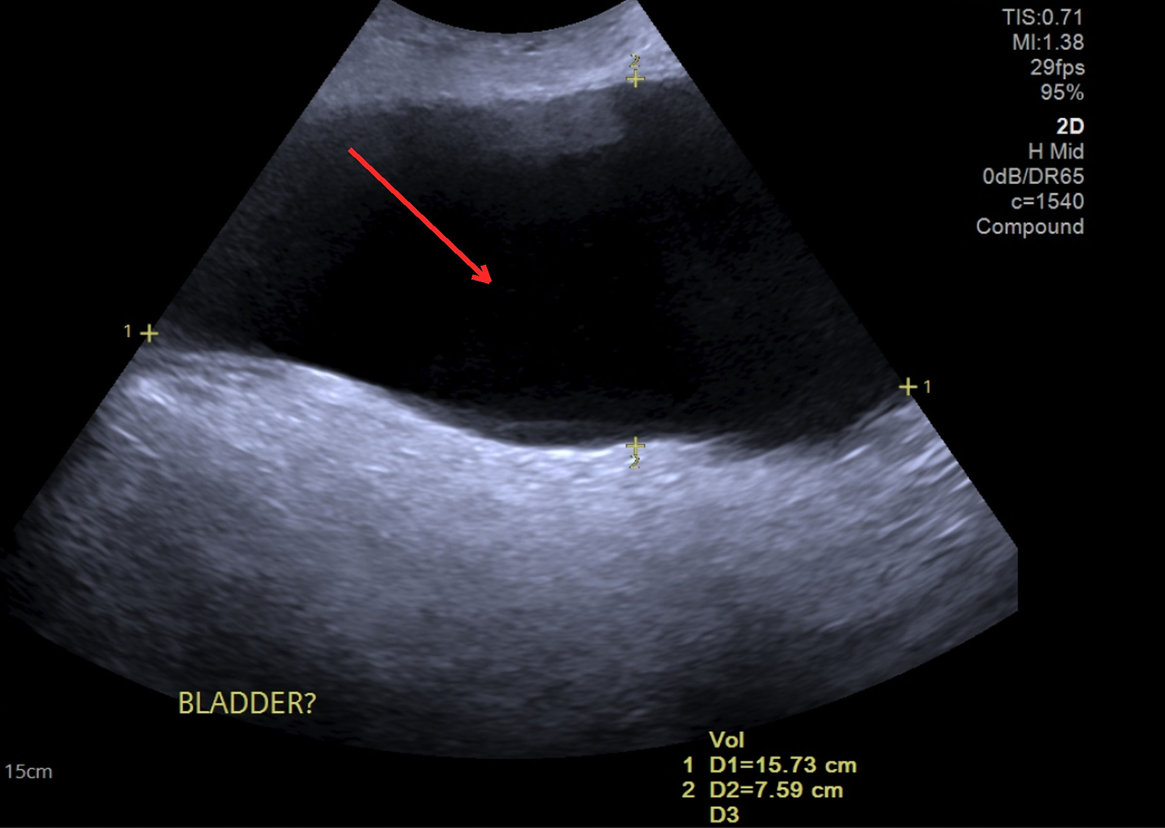
↓  Figure 1. Transabdominal and scrotal ultrasound. Large anechoic, fluid-filled structure exceeding 800 mL extending from the pelvis into the scrotum (red arrow), initially reported as a query bladder and in keeping with the herniated urinary bladder. Ultrasound was the first-line study and raised the initial suspicion of bladder herniation, before cross-sectional imaging was obtained.
Figure 1.
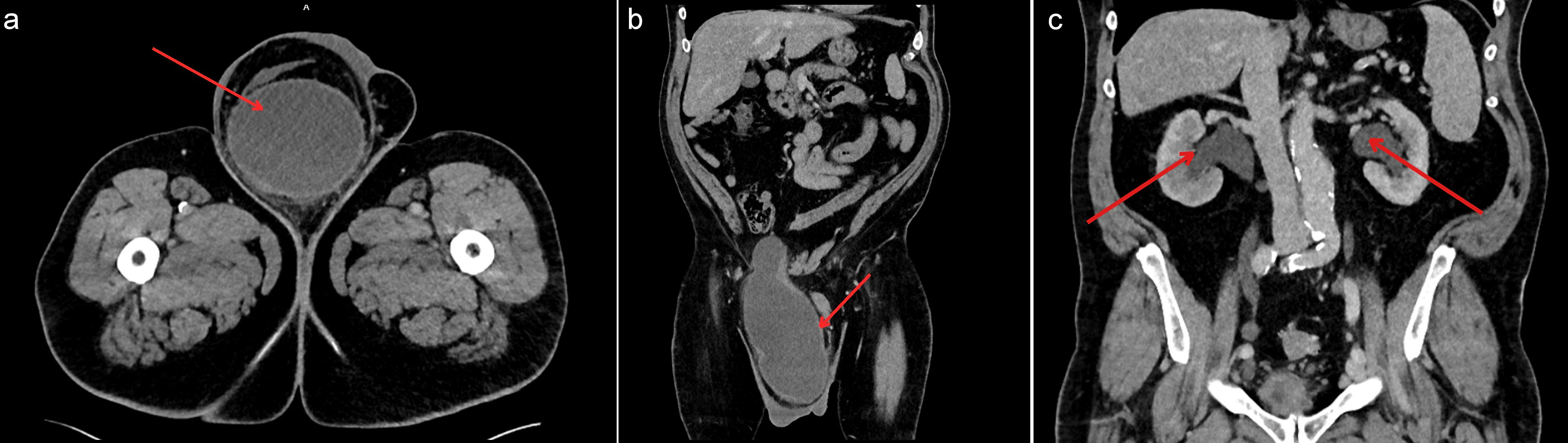
↓  Figure 2. Contrast-enhanced CT of the abdomen and pelvis. (a) Axial image showing a large right inguinoscrotal hernia containing the majority of the urinary bladder. (b) Coronal image showing descent of the bladder from the pelvis into the right scrotum. (c) Coronal image at the level of the kidneys demonstrating bilateral hydronephrosis with dilatation of the renal pelves and proximal ureters (red arrows indicate the dilated pelvicalyceal systems). In panels (a) and (b), the red arrow indicates the herniated bladder. CT: computed tomography.
Figure 2.

Table

↓  Table 1. Timeline of Clinical Events
 
Time pointEvent/finding
eGFR: estimated glomerular filtration rate; PSA: prostate-specific antigen; CT: computed tomography.
PresentationLongstanding right scrotal swelling; nocturnal leakage; obligatory seated voiding; urgency and frequency. eGFR 78 mL/min/1.73 m2, creatinine 86 µmol/L; PSA normal.
ImagingUltrasound: pelvic fluid collection > 800 mL extending into the scrotum (kidneys not imaged). Contrast-enhanced CT: right inguinoscrotal hernia containing most of the bladder with bilateral ureteric compression and hydronephrosis.
DeteriorationRapid renal decline (2 days after the creatinine of 86 µmol/L): eGFR 48 mL/min/1.73 m2, creatinine 126 µmol/L (acute kidney injury).
CatheterizationUrethral catheter drained 500–600 mL with immediate decompression and marked reduction in scrotal swelling.
SurgeryUrgent open right inguinal repair: coexisting direct and indirect hernias, bladder within the direct component; reduction without resection, posterior wall reconstruction, polypropylene mesh, intraoperative cystography confirming integrity.
PostoperativeSelf-limiting hematuria on day 1; catheter for 5 days; discharged on day 2.
2 weeksFlexible cystoscopy excluded intravesical pathology; successful trial without catheter.
8 monthsRenal function normalized (eGFR > 90 mL/min/1.73 m2); no recurrence or urinary complications.