| World Journal of Nephrology and Urology, ISSN 1927-1239 print, 1927-1247 online, Open Access |
| Article copyright, the authors; Journal compilation copyright, World J Nephrol Urol and Elmer Press Inc |
| Journal website https://wjnu.elmerpub.com |
Case Report
Volume 000, Number 000, August 2026, pages 000-000

Organ-Preserving Chemoradiotherapy and Successful Metastasectomy in a Rare Epithelioid Leiomyosarcoma of the Prostate
Arjun Nagaraja, Bhavyashree Shettya, b, Dinesh Annea, Murali Krishnaa, Abhishek Pasama
aDepartment of Urology, Sapthagiri Super Speciality Hospital, Hesaraghatta, Bangalore, India
bCorresponding Author: Bhavyashree Shetty, Department of Urology, Sapthagiri Super Speciality Hospital, Hesaraghatta, Bangalore, India
Manuscript submitted June 3, 2026, accepted August 7, 2026, published online August 21, 2026
Short title: Chemoradiotherapy and Metastasectomy in Prostate
doi: https://doi.org/10.14740/wjnu1058
| Abstract | ▴Top |
Epithelioid leiomyosarcoma is an aggressive soft tissue sarcoma derived from smooth muscle cells, typically arising from uterine, gastrointestinal, or deep soft tissue sites. Its primary occurrence in the prostate is rare, accounting for less than 0.1% of primary prostate malignancies. We present a case report of a 46-year-old male who presented with burning micturition, intermittent hematuria, and pelvic pain as his primary concerns. He had a cystic–solid mass centered at the left prostatic–urethral junction. Imaging studies, including ultrasound, contrast-enhanced computed tomography (CT), magnetic resonance imaging (MRI), and positron emission tomography-CT (PET-CT), revealed a heterogeneous lesion abutting the bladder base and the rectum without overt invasion. Transrectal biopsy with immunohistochemistry confirmed high-grade epithelioid leiomyosarcoma of the prostate, with tumor cells positive for vimentin, smooth muscle actin, calponin, desmin, and H-caldesmon, and negative for cytokeratin 5/6, P63, melanocytic and vascular markers. These findings were consistent with smooth muscle differentiation, effectively ruling out sarcomatoid carcinoma and melanoma. The patient received neoadjuvant chemotherapy with ifosfamide and doxorubicin, followed by definitive external beam radiotherapy with volumetric modulated arc therapy (VMAT). The treatment achieved a complete metabolic response, as evidenced by post-treatment PET-CT without the need for radical surgery. At 1-year follow-up, the patient presented with acute intestinal obstruction and jejunal leiomyosarcoma (grade 2) with a high proliferative index. Staging PET-CT also demonstrated a right lung nodule, which was confirmed cytologically as metastatic spindle cell sarcoma. He was treated with gemcitabine–docetaxel chemotherapy and video-assisted thoracoscopic right lung lobectomy. He remained well at the most recent follow-up with no clinical evidence of recurrent or metastatic disease. This case is unique for demonstrating that primary epithelioid leiomyosarcoma of the prostate can achieve a complete metabolic response with chemoradiotherapy alone, avoiding upfront radical prostatectomy, an organ-preserving outcome rarely described for this tumor, even after later metachronous jejunal and pulmonary metastases, which were successfully controlled with systemic therapy and metastasectomy. It underscores the aggressive metastatic behavior of prostatic leiomyosarcoma, the potential role of neoadjuvant chemotherapy and radiotherapy in organ preservation, and the value of ongoing surveillance for late, metachronous metastatic disease. The key learning points include recognizing atypical presentations of prostatic sarcoma, the role of multimodal therapy for local control, and individualizing surgical management of metachronous metastases.
Keywords: Chemoradiotherapy; Epithelioid leiomyosarcoma; Jejunal leiomyosarcoma; Metachronous metastasis; Prostate; Pulmonary metastasis; Sarcoma; Immunohistochemistry
| Introduction | ▴Top |
Sarcomas are malignant tumors arising from mesenchymal cell lines and comprise a heterogeneous group of cancers with distinct clinical, histologic, and radiographic characteristics [1]. Of all soft tissue sarcomas, approximately 10–20% are leiomyosarcomas, which typically arise in the uterus, retroperitoneum, gastrointestinal tract, or deep soft tissues. Primary leiomyosarcoma of the prostate is extremely rare, accounting for less than 0.1% of primary prostate malignancies and approximately 38–52% of primary prostatic sarcomas in adults [2, 3].
Clinically, prostatic leiomyosarcomas often present with lower urinary tract symptoms such as obstructive voiding, hematuria, pelvic pain, and occasionally urinary retention, and show prostate-specific antigen (PSA) levels that are typically within the normal range [2, 3].
We describe a case of epithelioid leiomyosarcoma of the prostate achieving a complete metabolic response to neoadjuvant chemoradiotherapy, without upfront radical prostatectomy, followed by metachronous jejunal and pulmonary metastases successfully controlled with systemic therapy and metastasectomy. This case adds to the limited literature on epithelioid variants and highlights the role of multimodal therapy and long-term surveillance.
| Case Report | ▴Top |
Investigations (patient information and clinical findings)
A 46-year-old male presented with a 1-week history of burning micturition, two episodes of hematuria, and pain in the pelvic region. There were no symptoms of hesitancy, urinary frequency, or urgency. There was no significant past medical history, family history of malignancy, or prior surgical interventions. The patient had no known psychosocial history of relevance and was not on any regular medications.
On rectal examination, a mass was palpated approximately 4 cm from the anal verge, which was cystic to firm in consistency. Baseline hematological and biochemical parameters were within normal limits. The PSA value was 4.04 ng/mL, which was marginally above the conventional upper reference threshold of 4 ng/mL.
Transabdominal pelvic ultrasound demonstrated an irregular, heterogeneously enhancing lesion adjacent to the lateral aspect of the prostate and the left seminal vesicle, suggestive of a neoplastic process, such as a seminal vesicle mass or a stromal tumor of the prostate. The spiral computed tomography of the kidneys, ureters, and bladder (CT KUB) showed an irregular, heterogeneously attenuating lesion predominantly involving the left lateral aspect of the prostate gland and left seminal vesicle, measuring 47 × 47 × 50 mm (craniocaudal × transverse × anteroposterior). The epicenter of the lesion was at the junction of the left prostate and the seminal vesicle. The lesion indented the rectum posteriorly and the bladder base anteriorly with loss of the intervening fat plane.
Magnetic resonance imaging (MRI) of the pelvis revealed an irregular mixed-signal intensity lesion in relation to the lateral aspect of the prostate and seminal vesicle region measuring 4.6 × 4.8 × 4.8 cm. The lesion showed diffuse diffusion restriction and heterogeneous post-contrast enhancement, with an enhancing peripheral solid component and central non-enhancing cystic/necrotic areas. The epicenter was confirmed at the left prostatic–seminal vesicle junction. The lesion distorted the prostate–seminal vesicle angle, displaced the left neurovascular bundle, and indented the rectum and bladder base without clear evidence of invasion.
Fluorine-18 fluorodeoxyglucose positron emission tomography-CT (18F-FDG PET-CT) was performed for further characterization and staging. It showed a 6.2 × 4.5 × 5.5 cm peripherally enhancing necrotic lesion involving the posterolateral and central glands of the prostate, with significant indentation of the seminal vesicles and anterior rectal wall, and mild indentation of the bladder base (Fig. 1). A peripheral rim of FDG uptake was noted in the gland, and the initial impression favored an inflammatory mass rather than a definite neoplasm. No distant FDG-avid lesions were identified at that time.
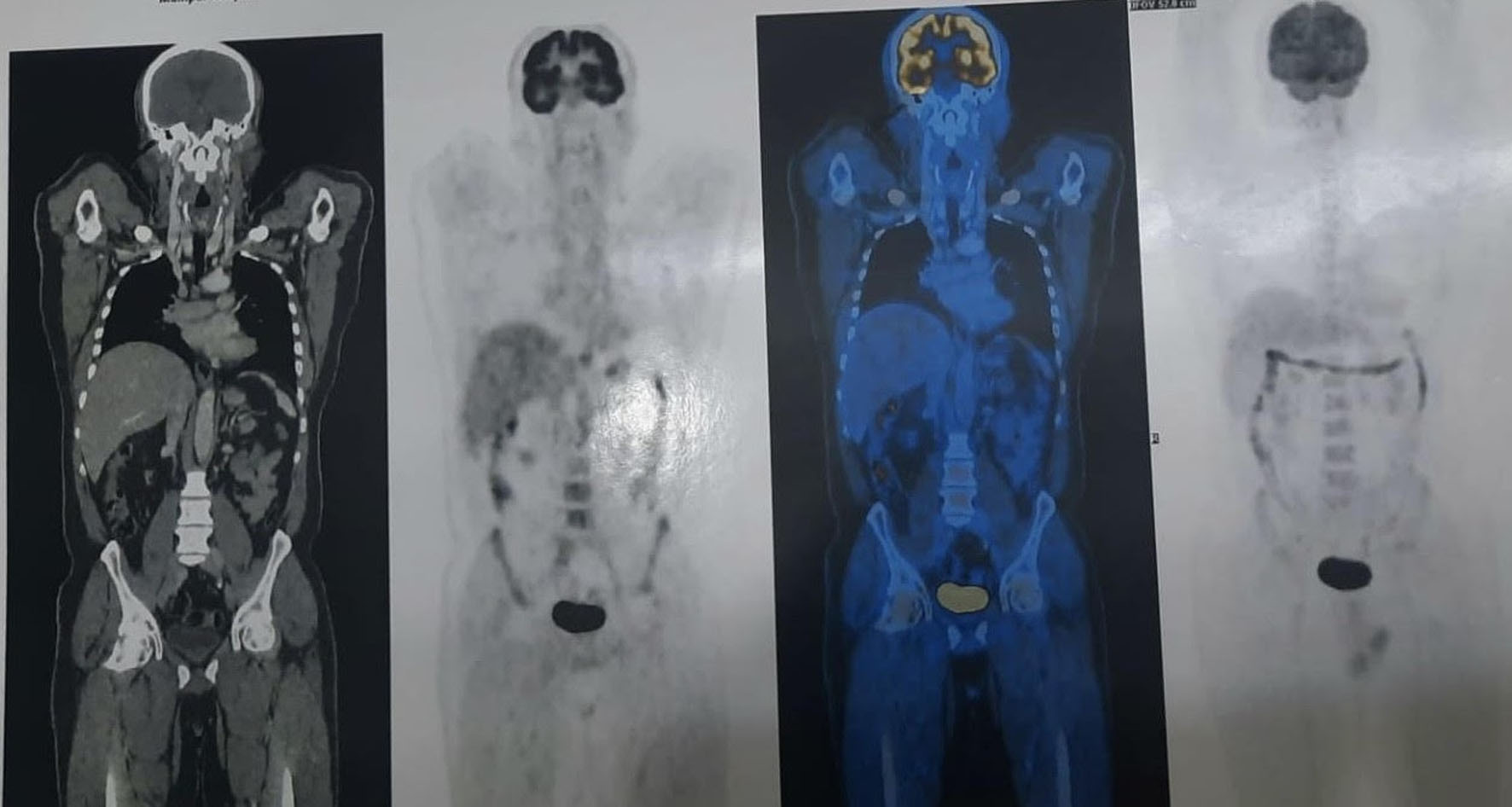 Click for large image | Figure 1. A peripherally FDG-avid, centrally necrotic lesion (6.2 × 4.5 × 5.5 cm) is seen involving the posterolateral and central glands of the prostate, with significant indentation of the seminal vesicles and anterior rectal wall, and mild indentation of the bladder base. A peripheral rim of FDG uptake is noted without focal hypermetabolic activity in the central necrotic zone. No distant FDG-avid lesions are identified, consistent with localised disease at initial staging. FDG: fluorodeoxyglucose. |
The timeline of the clinical course is presented in Table 1.
 Click to view | Table 1. Timeline of Clinical Course |
Diagnosis
Cystoscopy demonstrated non-obstructive prostatic lobes with normal bladder mucosa; both ureteric orifices were normal. Transrectal biopsy of the mass was performed, and fluid aspirated from the cystic portion was sent for cytology. Histopathological examination revealed a high-grade sarcoma composed of epithelioid cells consistent with epithelioid leiomyosarcoma (Figs 2 and 3). The results of immunohistochemical analysis are presented in Table 2.
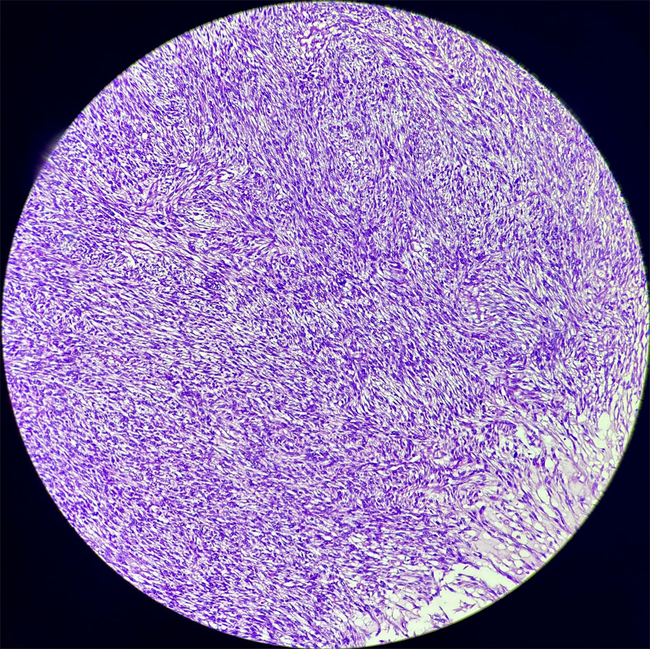 Click for large image | Figure 2. Section showing malignant oval to spindle cells arranged in fascicles and sheets. The cells exhibit a moderate degree of pleomorphism with coarse, clumped chromatin; occasional cells display bizarre nuclei. These morphological features, in conjunction with the immunohistochemical profile (SMA, desmin, calponin, H-caldesmon positive; epithelial and melanocytic markers negative), are consistent with high-grade epithelioid leiomyosarcoma of the prostate. H&E stain, low power. H&E: hematoxylin and eosin; SMA: smooth muscle actin. |
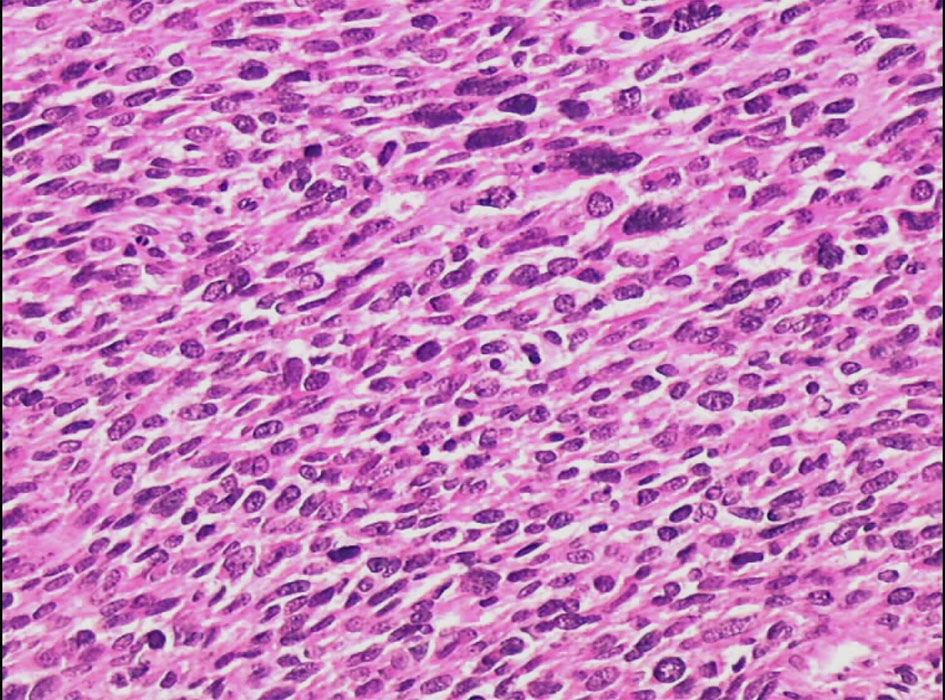 Click for large image | Figure 3. Spindle-shaped tumor cells arranged in interlacing fascicles with focal whorled patterns, characteristic of leiomyosarcoma. Marked nuclear pleomorphism, hyperchromasia, and increased mitotic activity are evident. This high-power view highlights the smooth muscle differentiation pattern consistent with a diagnosis of leiomyosarcoma of the prostate. H&E stain, high power. |
Click to view | Table 2. Immunohistochemical Analysis |
The combination of positive smooth muscle markers (smooth muscle actin (SMA), calponin, desmin, H-caldesmon) supported a diagnosis of smooth muscle tumor, while negative vascular (CD31, CD34, D2-40, ERG) and melanocytic markers (HMB45, melan-A, S-100) excluded angiosarcoma and malignant melanoma. Negative basal cell and squamous markers (CK5/6, P63, 34βE12) and epithelial membrane antigen (EMA) helped differentiate this lesion from sarcomatoid carcinoma and other spindle cell epithelial neoplasms of the prostate. The main differential diagnoses considered were sarcomatoid carcinoma, prostatic stromal sarcoma (stromal tumor of uncertain malignant potential (STUMP)), rhabdomyosarcoma, and angiosarcoma, all of which were excluded based on the immunohistochemical profile summarized in Table 2.
Based on morphology and immunophenotype, a final diagnosis of high-grade epithelioid leiomyosarcoma of the prostate was established.
Treatment
Given the high-grade nature and locally advanced disease abutting critical pelvic structures, a multidisciplinary tumor board recommended an organ-preserving strategy, that is, neoadjuvant chemotherapy followed by definitive radiotherapy with curative intent, with surgery reserved for non-responders or residual disease. This is consistent with the evidence that multimodal therapy may improve local control and survival in prostatic leiomyosarcoma.
The patient received neoadjuvant chemotherapy with ifosfamide (3.3 g on days 1–4) and adriamycin (doxorubicin, 120 mg as a continuous infusion over 72 h) in combination with pegylated granulocyte-colony stimulating factor (G-CSF) 6 mg for primary prophylaxis. After two cycles, interim PET-CT showed a good metabolic response of the prostatic lesion. Two additional cycles (third and fourth) of the same regimen were administered.
Subsequently, he underwent definitive external beam radiotherapy to the prostate and peri-prostatic region using a linear accelerator (LINAC) with VMAT technique. Three gold seed fiducial markers were placed in the prostate for image-guided radiotherapy. A total dose equivalent to standard radical prostate radiotherapy (76 Gy in conventional fractions over 7.5 weeks) was delivered, with careful sparing of surrounding organs at risk.
Post-chemoradiotherapy assessment included clinical examination and repeat PET-CT, which revealed no abnormal FDG uptake in the prostate gland and no metabolically active lesions elsewhere, consistent with a complete metabolic response.
Follow-up and outcomes
The patient was followed regularly with clinical evaluation and interval imaging. Nearly 12 months after the completion of chemoradiotherapy, in April 2021, he presented with acute intestinal obstruction and underwent emergency exploratory laparotomy. Intraoperatively, a segmental jejunal mass was identified and resected with primary anastomosis. Histopathology confirmed leiomyosarcoma of the jejunum (grade 2), with SMA diffusely positive, H-caldesmon positive, and a markedly elevated Ki-67 proliferative index of 60–70%, consistent with a high-grade smooth muscle sarcoma. Jejunal leiomyosarcoma itself is rare, further underscoring the aggressive and multifocal potential of leiomyosarcoma in this patient [4, 5].
A staging PET-CT performed during the same admission identified a right lung nodule. Fine-needle aspiration cytology (FNAC) from the lung lesion showed atypical spindle cells suggestive of metastatic sarcoma. The patient received four cycles of palliative-intent chemotherapy with gemcitabine (2,000 mg) and docetaxel (80 mg), a regimen commonly used for soft tissue sarcomas, including leiomyosarcoma. Following radiological reassessment and a favorable response, he underwent video-assisted thoracoscopic surgery (VATS) with right lung lobectomy.
PET-CT was done 6 months after metastasectomy, which showed no abnormal FDG uptake in the prostate gland, indicating a sustained complete metabolic response, and no local prostatic recurrence. At the most recent follow-up, the patient was doing well, performing routine daily activities with good performance status.
| Discussion | ▴Top |
Leiomyosarcomas represent a small fraction of soft-tissue sarcomas overall but are the most common primary sarcomas of the adult prostate [3, 6]. Prostatic leiomyosarcoma usually presents in middle-aged to older men with lower urinary tract symptoms, hematuria, pelvic pain, or urinary retention [2, 3, 7]; up to one-third of patients have metastatic disease at initial presentation, most often to the lungs and occasionally to the liver and bone [3, 7]. In the present case, the patient’s initial symptoms were relatively mild (burning micturition, intermittent hematuria, pelvic discomfort), and imaging suggested a cystic–solid mass at the prostatic–seminal vesicle junction without overt distant disease, highlighting the diagnostic challenge and potential for misinterpretation as inflammatory or benign lesions on imaging [3, 7].
The histological spectrum of smooth muscle tumors of the prostate includes leiomyoma, atypical smooth muscle tumors, and leiomyosarcoma, as well as other spindle cell lesions such as inflammatory myofibroblastic tumor, rhabdomyosarcoma, gastrointestinal stromal tumor (GIST), sarcomatoid carcinoma, STUMP, and prostatic stromal sarcoma [7–9]. Hansel et al (2007) emphasized the importance of morphology and a targeted immunohistochemical panel for distinguishing these entities, as their management and prognosis differ markedly [8]. In our case, positivity for vimentin, SMA, calponin, desmin, and H-caldesmon [6–8] and negativity for epithelial, melanocytic, and vascular markers [7] supported a diagnosis of leiomyosarcoma and effectively excluded sarcomatoid carcinoma, melanoma, and vascular neoplasms.
Epithelioid leiomyosarcoma is characterized by round-to-polygonal cells with eosinophilic or clear cytoplasm, round nuclei [10] with delicate chromatin, and occasional prominent nucleoli; the cells often form nests, cords, or sheets, sometimes with stromal hyalinization. Such phenotypic features may mimic carcinoma or other epithelioid neoplasms, making immunohistochemistry crucial in establishing the diagnosis [8]. The relatively high Ki-67 index (30–40% in the primary lesion and 60–70% in the jejunal lesion) in our patient is consistent with the aggressive behavior reported in the literature [10, 11].
There are no standardized treatment guidelines for primary prostatic leiomyosarcoma due to its rarity [7]. Most published reports describe multimodal approaches combining radical surgery, chemotherapy, and radiotherapy [2, 7]. Cheville et al (1995) reported a poor overall prognosis in a series of 23 cases, with many patients dying within a few years despite aggressive local therapy [2]. Another review similarly highlighted variable but generally unfavorable outcomes and suggested that negative surgical margins, absence of metastases at diagnosis, and use of multimodal therapy may improve survival [7]. More recent reports, including gemcitabine–docetaxel–based regimens and combined chemoradiotherapy, have demonstrated complete or durable responses in selected patients, as seen in our case [12].
Unlike many published studies, in which radical surgery has been the mainstay of local treatment [2, 7], our patient’s initial management with neoadjuvant ifosfamide–doxorubicin chemotherapy followed by high-dose, image-guided VMAT-based radiotherapy achieved a complete metabolic response without upfront radical prostatectomy. To our knowledge, complete response to chemoradiotherapy, avoiding radical prostatectomy, has rarely been reported for primary prostatic leiomyosarcoma, underscoring the novelty of this organ-preserving outcome. This organ-preserving approach may be considered in selected patients where radical pelvic surgery would be morbid and where a good response to systemic therapy is anticipated. However, evidence remains limited and such decisions must be individualized.
The subsequent development of metachronous jejunal and pulmonary metastases, after 12 months of completing chemoradiotherapy, underscores the high metastatic potential of leiomyosarcoma, particularly to the lung, liver, bone [7], and intra-abdominal sites. Jejunal leiomyosarcoma itself is rare, and gastrointestinal involvement in patients with a history of extraintestinal leiomyosarcoma is infrequently reported [4]. In this case, the combination of systemic chemotherapy with gemcitabine–docetaxel followed by surgical metastasectomy (jejunal resection and right lung lobectomy) resulted in good disease control [12] and functional outcomes, aligning with the broader sarcoma literature, in which resection of isolated or limited lung metastases can confer survival benefit in carefully selected patients [13].
Strengths and limitations
The strengths of this case include comprehensive multimodality imaging, detailed histopathological and immunohistochemical characterization, and documentation of longitudinal outcomes, including metachronous metastases and their successful management. Limitations include the single-patient nature of the report, the lack of molecular profiling, and the limited follow-up duration, which precludes firm conclusions about long-term survival. Nonetheless, the case contributes valuable information on the epithelioid variant of prostatic leiomyosarcoma, the feasibility of chemoradiotherapy-based organ preservation, and the role of metastasectomy in metachronous metastatic disease.
Learning points
Epithelioid leiomyosarcoma of the prostate is an exceptionally rare and aggressive malignancy that may present with non-specific urinary symptoms and can be radiologically misleading; a combination of histology and focused immunohistochemistry is essential for accurate diagnosis. Multimodal therapy, including neoadjuvant chemotherapy and high-precision radiotherapy, can achieve a complete response in selected patients and may allow avoidance of morbid radical pelvic surgery. Given the high propensity for hematogenous spread and the possibility of metachronous metastases to sites such as the jejunum and lung, long-term surveillance is crucial, and surgical metastasectomy alongside systemic therapy should be considered in fit patients with resectable lesions.
Acknowledgments
The authors also gratefully acknowledge the contributions of the multidisciplinary team—including urology, medical oncology, radiation oncology, pathology, and diagnostic radiology —at Sapthagiri Super Speciality Hospital, Bengaluru, whose collective expertise was central to the clinical management of this patient. The authors additionally acknowledge Dr Ruchi Agrawal (Medical Writer) for assistance with literature review and manuscript preparation.
Financial Disclosure
No specific grant from any funding agency in the public, commercial, or not-for-profit sectors was received for the conduct of this work or the preparation of this manuscript.
Conflict of Interest
The authors have no financial, personal, or professional conflicts of interest related to the subject matter or materials discussed in this case report.
Informed Consent
Written informed consent was obtained from the patient for publication of this case report, including all clinical details, imaging findings, histopathological images, and any other accompanying data, in accordance with the journal’s editorial policies and the CARE guidelines. Patient anonymity has been preserved throughout.
Author Contributions
Dr Arjun Nagaraj: concept and design; clinical management and decision-making; critical revision of the manuscript for medical content; final approval of the version to be published. Dr Bhavyashree Shetty (corresponding author): concept and design; data acquisition and clinical management; manuscript draft and revision; final approval of the version to be published. Dr Dinesh Anne: contribution in abstract and discussion section. Dr Murali Krishna: contribution in discussion and case report. Dr Abhishek Pasam: contribution in tables, references, and conclusion.
Data Availability
All relevant clinical, imaging, and histopathological data supporting the findings of this case report are contained within the article. No datasets were generated or analyzed beyond what is presented herein. Additional de-identified details may be made available upon reasonable request from the corresponding author (Dr Bhavyashree Shetty; drbhavyashetty@gmail.com), subject to institutional policies and patient confidentiality requirements.
Abbreviations
18F-FDG: fluorine-18 fluorodeoxyglucose; 34βE12: high molecular weight cytokeratin; CARE: case report guidelines; CD: cluster of differentiation; CK: cytokeratin; CK5/6: cytokeratin 5/6; CT: computed tomography; CT KUB: computed tomography of the kidneys, ureters, and bladder; D2-40: podoplanin (vascular marker); DRE: digital rectal examination; EMA: epithelial membrane antigen; ERG: ETS-related gene; FNAC: Fine-Needle Aspiration Cytology; G-CSF: granulocyte-colony stimulating factor; GIST: gastrointestinal stromal tumor; H&E: hematoxylin and eosin; H-caldesmon: heavy caldesmon; HMB-45: human melanoma black 45; IHC: immunohistochemistry; Ki-67: Kiel 67 proliferation index; LINAC: linear accelerator; melan-A: melanoma antigen A; MRI: magnetic resonance imaging; P63: tumor protein p63; PET-CT: positron emission tomography–computed tomography; PSA: prostate-specific antigen; S-100: S100 protein; SMA: smooth muscle actin; STUMP: stromal tumor of uncertain malignant potential; USG: ultrasonography; VATS: video-assisted thoracoscopic surgery; VMAT: volumetric modulated arc therapy
| References | ▴Top |
- Sbaraglia M, Bellan E, Dei Tos AP. The 2020 WHO classification of soft tissue tumours: news and perspectives. Pathologica. 2021;113(2):70-84.
doi pubmed - Cheville JC, Dundore PA, Nascimento AG, Meneses M, Kleer E, Farrow GM, Bostwick DG. Leiomyosarcoma of the prostate. Report of 23 cases. Cancer. 1995;76(8):1422-1427.
doi pubmed - Raj DH, Dash PK, Mohanty J, Sarangi PK. Leiomyosarcoma of the prostate-an unexpected histopathological outcome. BMJ Case Rep. 2016;2016:bcr2016215594.
doi pubmed - Elhence PA, Rao M, Vishnoi JR, Meena V. Jejunal leiomyosarcoma: report of a rare case diagnosed on cytology. Diagn Cytopathol. 2019;47(5):498-502.
doi pubmed - Aftan MK, Alfalahi A, Alzeena E, Albastaki U, Houcinat Y, Mahmoud K. Leiomyosarcoma: a rare presentation as multifocal lesion. BJR Case Rep. 2020;6(3):20190117.
doi pubmed - Mestiri S, Elghali MA, Bourigua R, Abdessayed N, Nasri S, Amine BA, Missaoui N, et al. Soft tissue leiomyosarcoma-diagnostics, management, and prognosis: data of the registry cancer of the center of Tunisia. Rare Tumors. 2019;11:2036361318820171.
doi pubmed - Venyo AK. A review of the literature on primary leiomyosarcoma of the prostate gland. Adv Urol. 2015;2015:485786.
doi pubmed - Hansel DE, Herawi M, Montgomery E, Epstein JI. Spindle cell lesions of the adult prostate. Mod Pathol. 2007;20(1):148-158.
doi pubmed - Marcal LP, Surabhi VR, Ramani NS, Katabathina VS, Paspulati RM, Prasad SR. Mesenchymal neoplasms of the prostate and seminal vesicles: spectrum of disease with radiologic-pathologic correlation. Radiographics. 2022;42(2):417-432.
doi pubmed - Chapel DB, Nucci MR, Quade BJ, Parra-Herran C. Epithelioid leiomyosarcoma of the uterus: modern outcome-based appraisal of diagnostic criteria in a large institutional series. Am J Surg Pathol. 2022;46(4):464-475.
doi pubmed - Mayerhofer K, Lozanov P, Bodner K, Bodner-Adler B, Obermair A, Kimberger O, Czerwenka K. Ki-67 and vascular endothelial growth factor expression in uterine leiomyosarcoma. Gynecol Oncol. 2004;92(1):175-179.
doi pubmed - Hensley ML, Maki R, Venkatraman E, Geller G, Lovegren M, Aghajanian C, Sabbatini P, et al. Gemcitabine and docetaxel in patients with unresectable leiomyosarcoma: results of a phase II trial. J Clin Oncol. 2002;20(12):2824-2831.
doi pubmed - Chang CH, Chiang XH, Lin MW, Kuo SW, Huang PM, Hsu HH, Chen JS. Outcome and survival analysis of pulmonary metastasectomy for primary sarcoma with pulmonary metastases. Front Surg. 2024;11:1470784.
doi pubmed
This article is distributed under the terms of the Creative Commons Attribution 4.0 International License (CC BY 4.0), which permits unrestricted use, distribution, and reproduction in any medium, including commercial use, provided the original work is properly cited.
World Journal of Nephrology and Urology is published by Elmer Press Inc.