World Journal of Nephrology and Urology, ISSN 1927-1239 print, 1927-1247 online, Open Access
Article copyright, the authors; Journal compilation copyright, World J Nephrol Urol and Elmer Press Inc
Journal website https://wjnu.elmerpub.com

Case Report

Volume 000, Number 000, August 2026, pages 000-000


Organ-Preserving Chemoradiotherapy and Successful Metastasectomy in a Rare Epithelioid Leiomyosarcoma of the Prostate

Figures

↓  Figure 1. A peripherally FDG-avid, centrally necrotic lesion (6.2 × 4.5 × 5.5 cm) is seen involving the posterolateral and central glands of the prostate, with significant indentation of the seminal vesicles and anterior rectal wall, and mild indentation of the bladder base. A peripheral rim of FDG uptake is noted without focal hypermetabolic activity in the central necrotic zone. No distant FDG-avid lesions are identified, consistent with localised disease at initial staging. FDG: fluorodeoxyglucose.
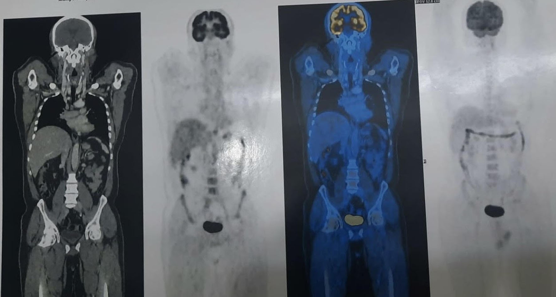
Figure 1.
↓  Figure 2. Section showing malignant oval to spindle cells arranged in fascicles and sheets. The cells exhibit a moderate degree of pleomorphism with coarse, clumped chromatin; occasional cells display bizarre nuclei. These morphological features, in conjunction with the immunohistochemical profile (SMA, desmin, calponin, H-caldesmon positive; epithelial and melanocytic markers negative), are consistent with high-grade epithelioid leiomyosarcoma of the prostate. H&E stain, low power. H&E: hematoxylin and eosin; SMA: smooth muscle actin.
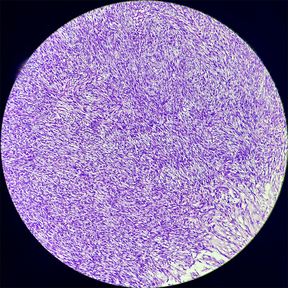
Figure 2.
↓  Figure 3. Spindle-shaped tumor cells arranged in interlacing fascicles with focal whorled patterns, characteristic of leiomyosarcoma. Marked nuclear pleomorphism, hyperchromasia, and increased mitotic activity are evident. This high-power view highlights the smooth muscle differentiation pattern consistent with a diagnosis of leiomyosarcoma of the prostate. H&E stain, high power.
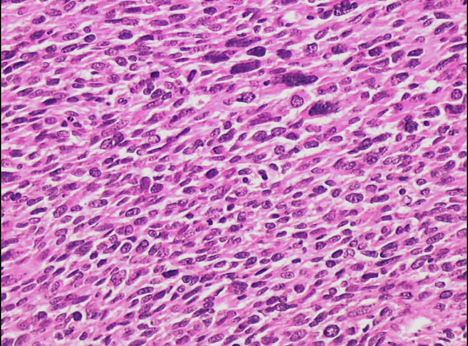
Figure 3.

Tables

↓  Table 1. Timeline of Clinical Course
 
Time pointEvent
DRE: digital rectal examination; USG: ultrasonography; CT KUB: computed tomography of the kidneys, ureters, and bladder; MRI: magnetic resonance imaging; PET-CT: positron emission tomography–computed tomography; VMAT: volumetric modulated arc therapy; VATS: video-assisted thoracoscopic surgery.
Initial presentationBurning micturition, haematuria, pelvic pain; DRE: cystic–firm mass at 4 cm from anal verge; normal labs.
Baseline imagingUSG, CT KUB, MRI pelvis: heterogeneous mass at left prostate–seminal vesicle junction with rectal and bladder indentation.
StagingPET-CT: peripherally enhancing necrotic lesion in prostate, interpreted as likely inflammatory mass; no distant disease.
Diagnostic procedureCystoscopy and transrectal biopsy with aspiration of cystic component; IHC confirming high-grade epithelioid leiomyosarcoma.
Primary treatmentNeoadjuvant ifosfamide–doxorubicin chemotherapy followed by VMAT-based external beam radiotherapy; complete metabolic response on PET-CT.
Metachronous eventApproximately one year later: acute intestinal obstruction; jejunal resection confirming grade 2 leiomyosarcoma; PET-CT revealed right lung nodule.
Management of metastasesGemcitabine–docetaxel chemotherapy followed by VATS right lung lobectomy; patient well on follow-up.


↓  Table 2. Immunohistochemical Analysis
 
MarkerResult
34βE12: high molecular weight cytokeratin; CD: cluster of differentiation; CK: cytokeratin; EMA: epithelial membrane antigen; ERG: ETS-related gene; H-caldesmon: heavy caldesmon; HMB45: human melanoma black 45; Ki-67: Kiel 67 proliferation index; melan-A: melanoma antigen A; P63: tumor protein p63; S-100: S100 protein; SMA: smooth muscle actin.
CKLocally positive
VimentinStrongly and diffusely positive
CalponinPositive
SMAPositive
DesminPositive
H-CaldesmonPositive (initial workup)
CK5/6, P63, EMA, 34βE12Negative
Vascular markers (CD31, CD34, D2-40, ERG)Negative
Melanocytic markers (HMB45 and Melan-A)Negative
S-100Negative
Ki-67 proliferation index30–40% in the primary prostate lesion