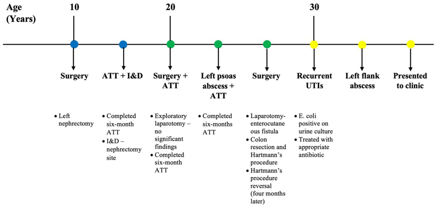
↓ Figure 1. Clinical timeline of the
patient’s course prior to presentation at our clinic, highlighting major interventions and
symptom progression. ATT: antitubercular therapy; E. coli: Escherichia coli; I&D:
incision and drainage; UTI: urinary tract infection.
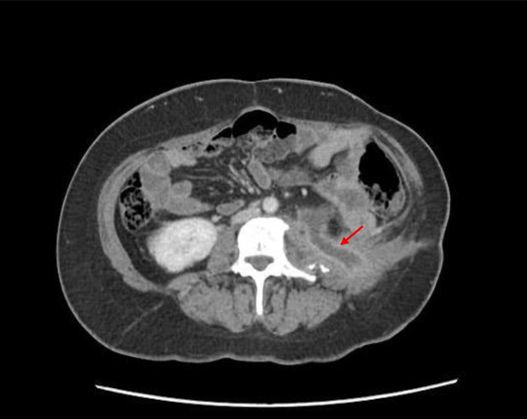
↓ Figure 2. Transverse plane of contrast-enhanced
computed tomography (CT) scan showing a cutaneous tract on the left side measuring approximately 28 mm
in length (arrow), along with evidence of prior left nephrectomy.
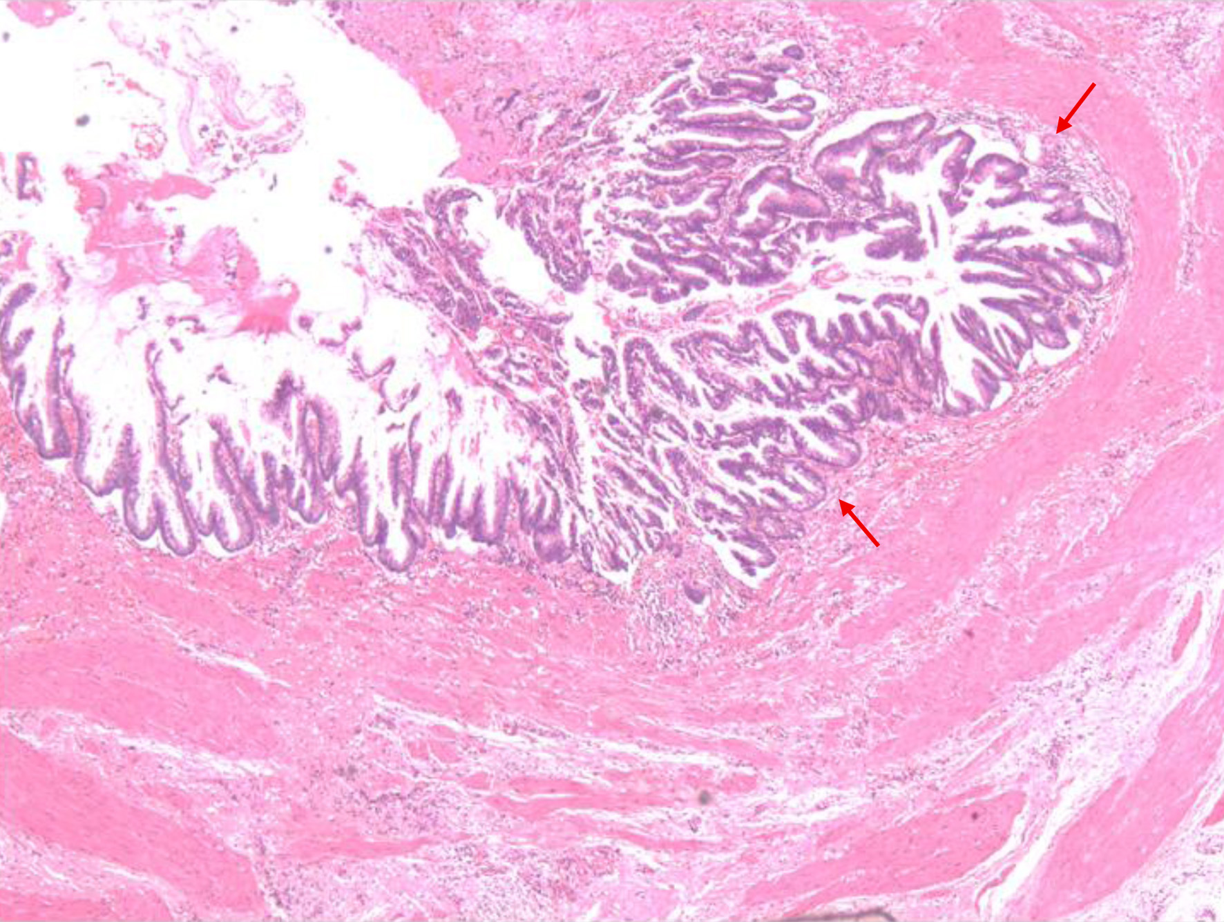
↓ Figure 4. Histopathological section of the
ureteric wall showing a lesion with papillary configuration, composed of fused tubules lined by columnar
epithelium and goblet cells exhibiting pseudostratification and low-grade dysplasia (arrows). No
evidence of muscularis or vascular invasion is identified.